C locus
C locus
The c locus, better known as the albino locus (c), is located on chromosome 7 of the mouse and is associated with mutations in the tyrosinase (Tyr) gene.23–28 It is important to note that albinism is epistatic (i.e., the situation in which the effect of one gene (phenotype) is dependent on the presence of one or more modifier genes) to all other coat color determinants; thus, all mice possessing a Tyrc/c genotype will lack pigment regardless of other coat alleles. This allele is the oldest allele that has been known and documented as far back as ancient Greek and Roman mouse fanciers.29,30 Interestingly, albino animals do not lack normal melanocytes, but the Tyrc group of alleles affects the amount of tyrosinase in the melanocyte. Tyrosinase is an enzyme that catalyzes the hydroxylation of L-tyrosine to
The human equivalent of the dilute locus is the MYO5A gene located at 15q21.12. Griscelli syndrome is an autosomal recessive disorder that is characterized by pigmentary dilution.46 Type 1, type 2, and type 3 Griscelli syndrome are distinguished by central nervous system involvement with hypopigmentation, immunological defects with hypopigmentation, and hypopigmentation alone, respectively.47,48 Type 3 is characterized by either homozygous deletion of the MYO5A F-exon, which is a tissue-specific exon that is transcribed in melanocytes, or by a homozygous mutation in the MLPH (melanophilin) gene.49
L-3,4-dihydroxyphenylalanine (L-dopa). L-dopa is then oxidized to dopaquinone, which is a common step in both the eumelanogenic and pheomelanogenic synthesis pathways.8 Once L-dopa is formed, melanogenesis can proceed further through oxidation-reduction reactions and spontaneous transformations. Although Tyr encodes for tyrosinase, some albino mutations affect tyrosinase activity rather than structure,31 introducing several other signaling pathways and genes that may affect albinism in mice.32 This is illustrated in Fig. 36.10, which shows that there are a number of genes that both directly and indirectly interact with Tyr. Normal appearing melanocytes can be found in the hair and eye, but they completely lack melanin. Pigment granules within the cells are significantly smaller and fewer than in wildtype genetic backgrounds.
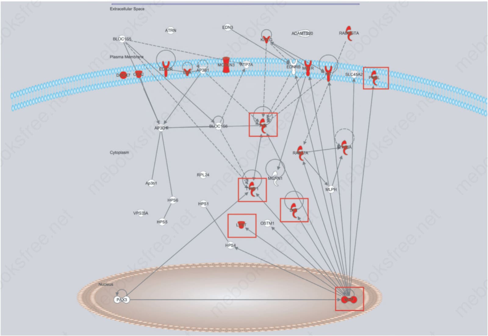
Fig. 36.10 Ingenuity Pathway Analysis Software® (http://www.ingenuity.com/) molecular pathway diagram for genes that interact in the coat color pathway. Many of the mutant mice in Fig. 36.9 have mutations in the genes in this pathway (red boxes). This illustrates how variations on a common trait, coat color, help to define a molecular pathway. It also illustrates the number of other genes involved with coat color not in this pathway, which will be future goals to integrate.