Perineurioma
Perineurioma
Clinical features Perineurioma is a neoplasm that was originally described as presenting in the subcutaneous tissue as soft tissue perineurioma or storiform perineural fibroma.1–4 However, the spectrum of perineurioma is wide and includes other variants such as cutaneous perineurioma, intraneural perineurioma
1802 Connective tissue tumors
(localized hypertrophic neuropathy) and sclerosing perineurioma.5–12 Perineurioma usually presents in adults and rarely in children.13 An association with neurofibromatosis has been described.14,15 Occurrence in the gastrointestinal tract, prostate, kidney, lung, oral cavity, parotid gland, as well as orbital and meningeal cases have been reported.16–33
• Cutaneous perineurioma is relatively common and presents as a small papular lesion, mainly on the lower limbs of middle-aged adults, with predilection for females.5–8 Behavior is benign with no tendency for local recurrence.
• Soft tissue perineurioma has very similar clinical features to those of cutaneous variants but lesions are subcutaneous and tend to be larger (up to 5 cm).1–4
• Intraneural perineurioma generally presents in young adults, and patients develop localized neurological symptoms as a result of intraneural growth.9,10,34 Multiple lesions and a case associated with amyotrophy have been reported.35,36
• Sclerosing perineurioma presents in young adults, with marked predilection for the fingers and palm. Extra-acral lesions are very rare.37–39 An exceptional case with bilateral lesions, and a patient with numerous tumors, have been documented.40,41 Behavior is benign.11,12
• Malignant perineuriomas are exceptional.42–48 Local recurrence has been reported but metastatic spread is rare.
Pathogenesis and histologic features Abnormalities of chromosome 22 and specifically deletion of NF2 have been reported in a few cases.10,49 In addition, 10q24 rearrangements and ABL1 gene involvement has been reported in two separate cases.50,51
Histologically, cutaneous perineurioma is a well-circumscribed, often dumbbell-shaped tumor composed of bland, short, spindle-shaped cells arranged in fascicles, with a focal whorling and a storiform pattern (Figs 35.370–35.372). Tumor cells may have focal epithelioid morphology. Variable hyalinization of the collagen is present and some cases show scattered, mononuclear inflammatory cells. Exceptional lesions with ossification, granular cell change and adipocytes or even lipoblasts have been described.52–55 An angiofibroma -like pattern has been reported.56 In a patient with multiple tumors, the lesions showed hybrid features of perineurioma and granular cell tumor.57 In a further case, granular cell change was documented.58 Soft tissue perineuriomas are very similar to those seen in the skin. Tumors are well circumscribed and generally cellular, being composed of monotonous bipolar cells with slender small nuclei in a fascicular, whorled or storiform growth pattern. Some cases are less cellular with a myxocollagenous stroma. Mitotic figures are rare and pleomorphism is absent.
A reticular variant of soft tissue perineurioma has been documented (Fig. 35.373).59–61 In this variant, a lace-like or reticular growth pattern composed
1803 Benign neural tumors
of anastomosing cords of spindle-shaped cells with pale pink cytoplasm and bipolar cytoplasmic processes is seen.
positive.64,69 In soft tissue perineurioma and sclerosing perineurioma, rare focal positivity for keratin has been described.11
Differential diagnosis Distinction from dermatofibrosarcoma protuberans is afforded by the latter’s diffuse CD34 positivity and infiltrative growth pattern. Other neural tumors usually lack a storiform pattern and are generally S100 protein positive.
In intraneural perineurioma, perineurial cells proliferate around individual axons with a characteristic onion ring appearance. A reticular pattern has been documented in one case.62,63
Sclerosing perineurioma is characterized by prominent hyalinized collagen around the tumor cells, which are arranged in cords, bundles and whorls (Figs 35.374 and 35.375). Trabecular, reticulated and whorled patterns may be seen.64 Cytologic atypia is absent and mitotic figures are exceptional. A single case contained mature adipocytes.65 Xanthomatous changes have been reported.66 A plexiform pattern and a whirling cellular variant are very rare.58,67,68
In all perineuriomas, tumor cells are diffusely positive for EMA, but negative for other neural markers (including S100 protein), in keeping with perineurial differentiation (Fig. 35.376).3,4 Focal and sometimes diffuse positivity for CD34 is seen in a small number of cases (Fig. 35.377) possibly reflecting the presence of fibroblasts. GLUT 1 and claudin-1 are usually
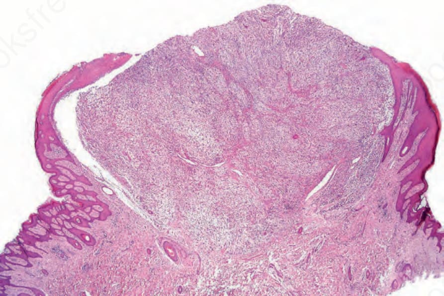
Fig. 35.370 Perineurioma: this tumor may closely resemble dermatofibrosarcoma protuberans but is generally better circumscribed.
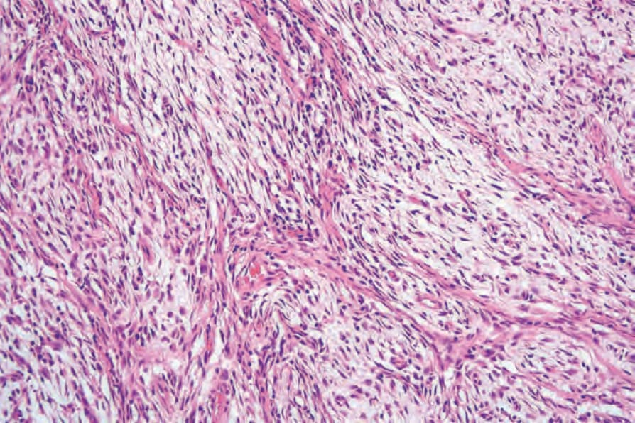
Fig. 35.371 Perineurioma: there is a whorled growth pattern.
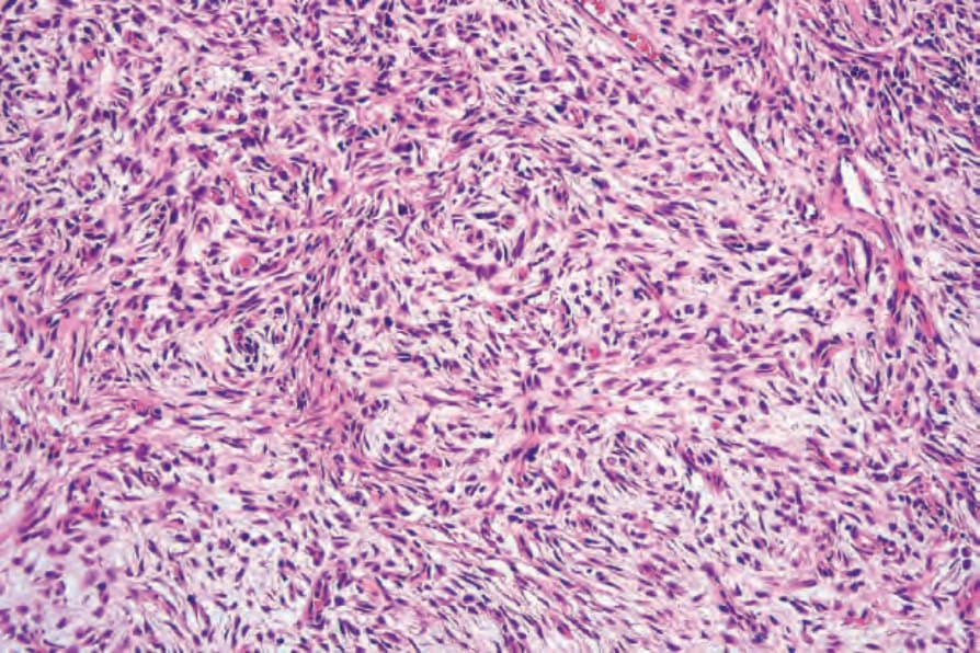
Fig. 35.372 Perineurioma: often, the tumor cells are arranged in a typical storiform growth pattern.
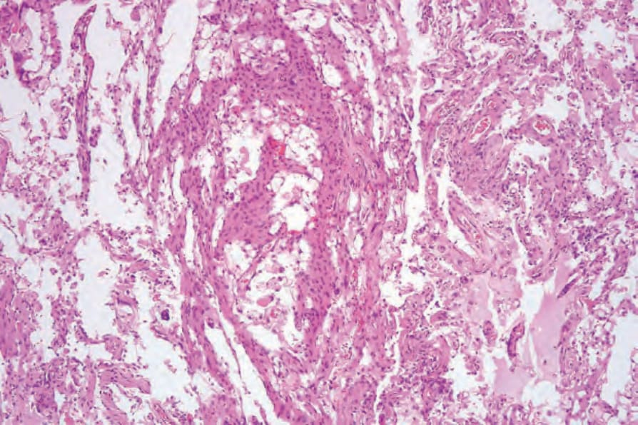
Fig. 35.373 Reticular perineurioma: note the lace-like growth pattern.
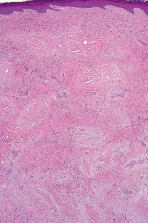
Fig. 35.374 Sclerosing perineurioma: this variant is characterized by a dense fibrous stroma.

Fig. 35.375 Sclerosing perineurioma: residual nodules are scattered throughout the lesion.
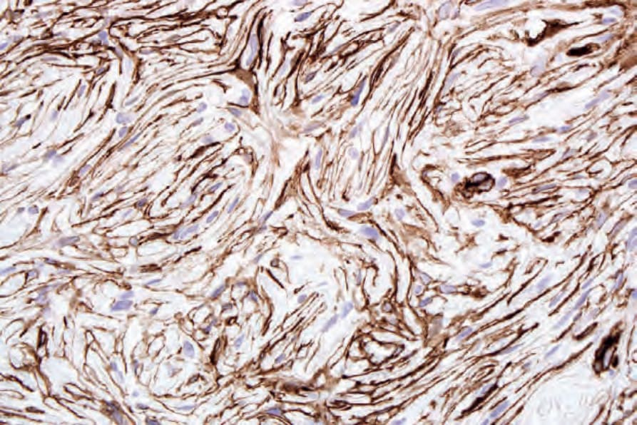
Fig. 35.376 Perineurioma: the tumor cells express epithelial membrane antigen.
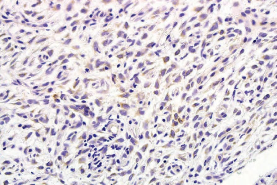
Fig. 35.377 Perineurioma: there is cytoplasmic CD34 expression.