Solitary circumscribed neuroma
Solitary circumscribed neuroma
Clinical features Solitary circumscribed neuroma (palisaded encapsulated neuroma) is a common but often unrecognized tumor that presents as a solitary,
1784 Connective tissue tumors
is identified near the base of the lesion, often entering (or fusing with) the lesional capsule. Prominent vascularity is occasionally present.20
By immunohistochemistry, most of the cells are S100 protein positive, in keeping with Schwann cells (Fig. 35.304). GFAP is negative.5 Numerous axons can be identified with neurofilament protein and the cells in the capsule stain for EMA, as expected in normal perineurial cells (Fig. 35.305).21,22 As the capsule tends to be partial and EMA can be weak, additional stains that help identifying the perineural cells include claudin 1 and Glut-1.5
Pathogenesis and histologic features The pathogenesis is unknown and there is no consensus as to whether the lesion is reactive or neoplastic.1–5 In a case associated with trauma, a hyperplastic phenomenon mediated by IL-6 was suggested.6
Histology characteristically shows fairly prominent nerves in the superficial dermis encased by cytologically bland squamous epithelium (Figs 35.306 and 35.307). There is no evidence of a connection to the overlying epidermis or neighboring adnexal structures. A loose myxoid stroma, a lymphocytic infiltrate and prominent infundibular cysts may be seen.4 Similar appearances may be seen in keratoacanthoma but in the latter there is evidence of inflammation and fibrosis accompanying the overlying tumor. In addition, the perineural invasion is usually deeply seated.
Differential diagnosis Distinction from neurofibroma and schwannoma is easy if attention is paid to the characteristic architecture and the presence of numerous intralesional axons.
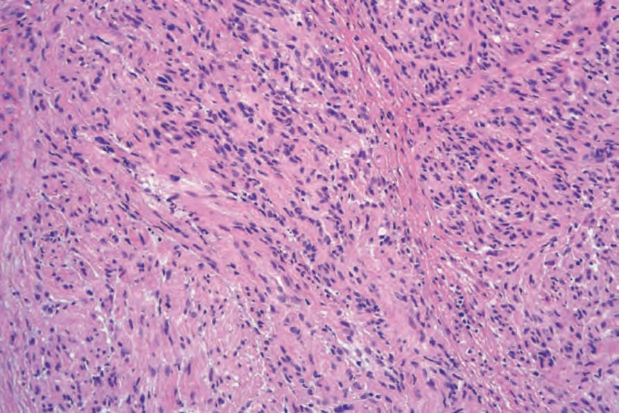
Fig. 35.301 Solitary circumscribed neuroma: medium-power view showing multinodularity.
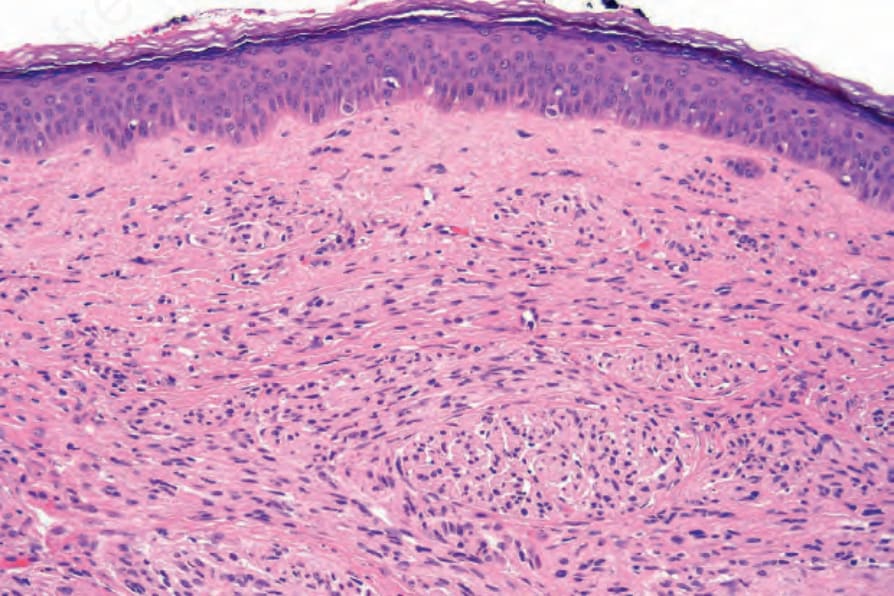
Fig. 35.302 Solitary circumscribed neuroma: note that the tumor merges imperceptibly into the papillary dermis.
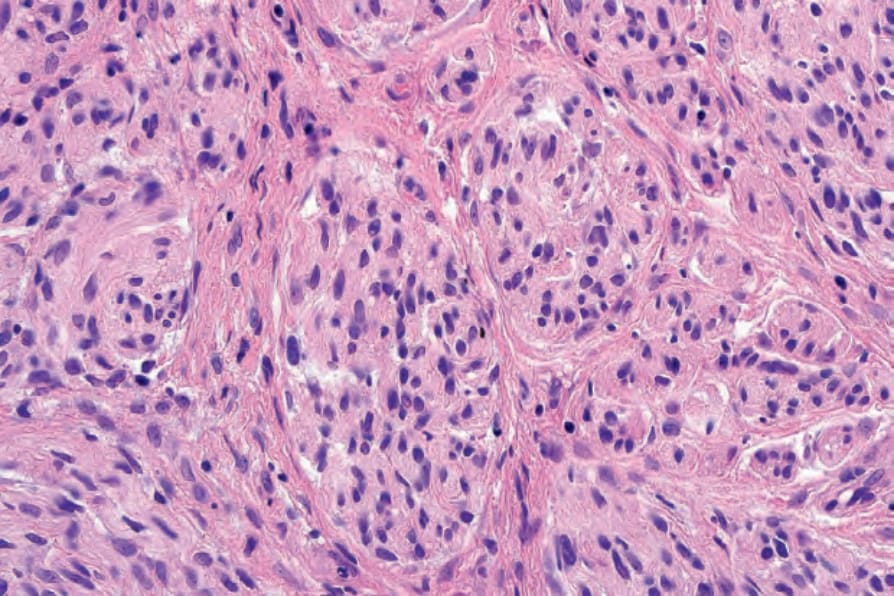
Fig. 35.303 Solitary circumscribed neuroma: the tumor is composed of pale-staining spindled cells with uniform elongated darkly staining nuclei.
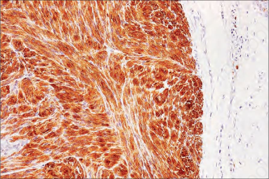
Fig. 35.304 Solitary circumscribed neuroma: the tumor cells express S100 protein.
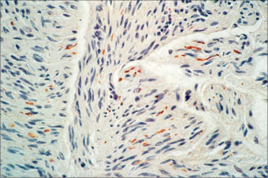
Fig. 35.305 Solitary circumscribed neuroma: the tumor contains numerous nerve fibers (neurofilament immunocytochemistry).
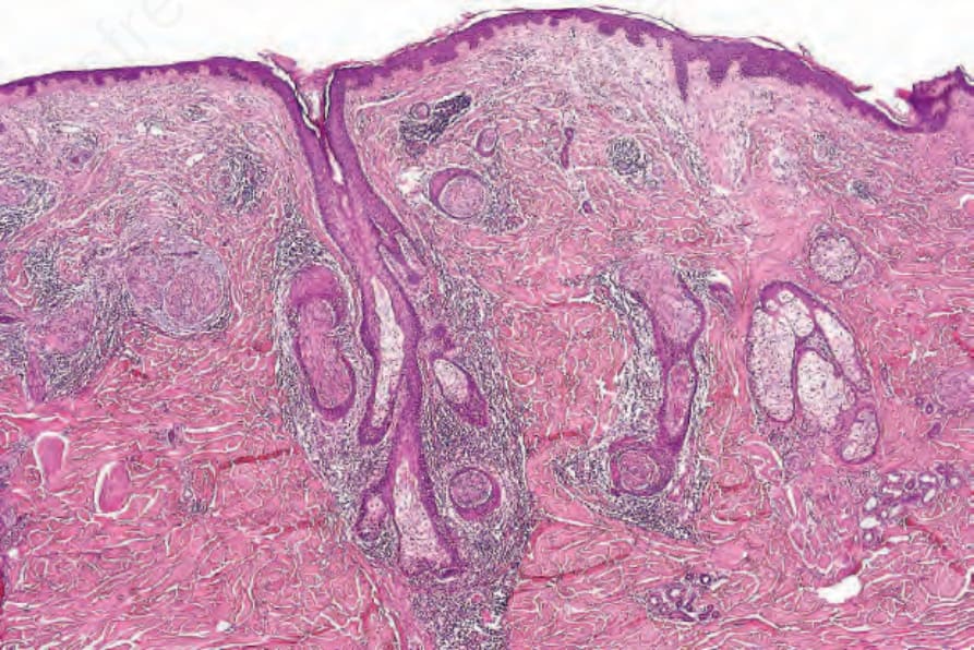
Fig. 35.306 Epithelial sheath neuroma: prominent nerves are present in the superficial reticular dermis encased by nests of bland squamous epithelium. By courtesy of L. Requena, MD, Madrid, Spain.