Myxofibrosarcoma
Myxofibrosarcoma
Clinical features Myxofibrosarcoma, formerly known as myxoid malignant fibrous histiocytoma is currently defined as a malignant fibroblastic tumor with pleomorphism, myxoid stroma and a distinctive curvilinear vascular pattern.1 The tumor commonly presents in the limbs of the elderly and shows a slight predilection for males.2–4 Up to 60% of cases arise in the subcutis and secondary involvement of the skin is common (Fig. 35.192).5–7 Prognosis
1756 Connective tissue tumors
is related to histologic grading, but behavior tends to be indolent with a high tendency for local recurrence, occasional metastasis to regional lymph nodes and a 5-year survival of up to 70%.4 Metastases are seen more commonly in deep-seated lesions and those with a high histologic grade.4 Local recurrences appear to be associated with higher histologic grade and more complex cytogenetic abnormalities, surgical margins (close or involved) and old age.8–12 Mortality is associated with tumor necrosis, large size and decrease in myxoid areas.13
Pathogenesis and histologic features Karyotypes are highly complex and no recurrent genetic changes have been established. In addition, correlation of molecular events with malignant potential has been studied in some series. Gains at chromosome 7 have been described, and more recently, overexpression of MET (chromosome 7q31) have been reported in myxofibrosarcoma and this feature is associated with deeper, higher-grade tumors in more advanced stages.14,15 A recent studies demonstrated mutations in TP53, ATRX, PTEN, FGFR3, CDKN2A, and RB1.16,17 MET is a transmembrane receptor tyrosine kinase representing the only high-affinity receptor of hepatocyte growth factor (HGF). Ezrin, as protein associated with cell adhesion-mediated signaling, is over-expressed in myxofibrosarcoma and the expression correlates with poor prognostic factors including necrosis, high mitotic activity, high histologic grade and advanced stage.18 Association of aggressiveness with down-regulation of p12CDK2AP1 has been reported.19 Of interest, chondroitin sulfate synthase 1 expression has been reported as associated with malignant potential in soft tissue sarcomas with myxoid substance.20 Domain-containing protein 2 (DCBLD2) is highly expressed in infiltrative myxofibrosarcoma.21
Histologically, appearances vary from low-grade, markedly myxoid, hypocellular lesions, to highly cellular, pleomorphic tumors with focal myxoid change. Tumor cells range from stellate to spindle shaped with variable pleomorphism. All tumors share a multinodular growth pattern, curvilinear thin-walled blood vessels, and a minimum of 10–20% of myxoid stroma with hyperchromatic stellate or spindle-shaped cells (Figs 35.193–35.198). Epithelioid cell change can be prominent, particularly in high-grade tumors.22
Tumor cells have ultrastructural features of fibroblasts and myofibroblasts and are only rarely focally for actin.4,23,24
Differential diagnosis Distinction from superficial angiomyxoma is easy because the latter lacks cytologic atypia, is less cellular, is predominantly dermal and commonly has
1757 Benign fibrohistiocytic tumors and tumorlike lesions
an epithelial component. Rare tumors can show focal changes mimicking a pleomorphic hyalinizing angiectatic tumor and sampling is very important to avoid a misdiagnosis.25 In low-grade fibromyxoid sarcoma, there is no pleomorphism, mitotic figures are rare and curvilinear blood vessels are usually absent.26 Immunohistochemical markers that have been reported to be of value in differential diagnosis from other myxoid soft tissue tumors include Claudin 6 and NY-ESO-1.27,28 The former is expressed in myxofibrosarcoma and the latter is expressed in myxoid/round cell liposarcoma but not in myxofibrosarcoma.
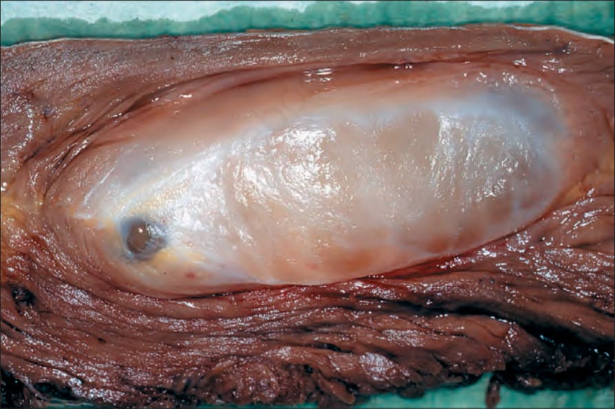
Fig. 35.192 Low-grade myxofibrosarcoma: note the multilobularity and prominent myxoid change. By courtesy of C.D.M. Fletcher, MD, Brigham and Women’s Hospital and Harvard Medical School, Boston, USA.
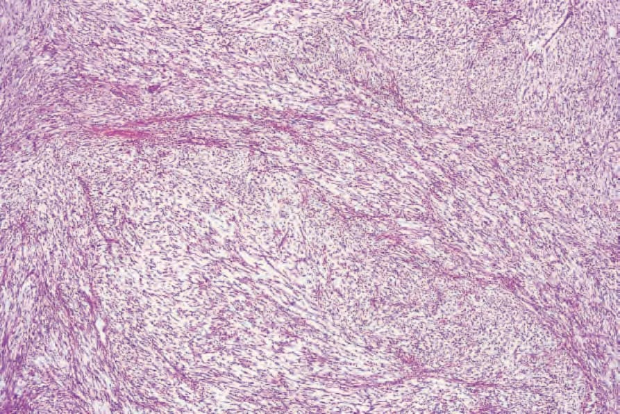
Fig. 35.193 Myxofibrosarcoma: low-grade lesions are relatively hypocellular and contain distinctive curvilinear vessels.
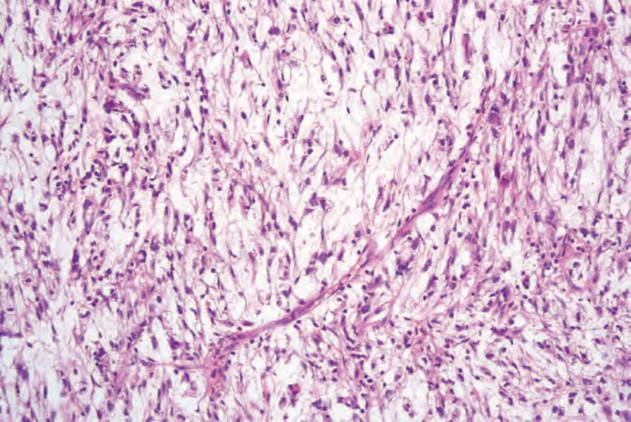
Fig. 35.194 Myxofibrosarcoma: the curvilinear vessels are characteristic.
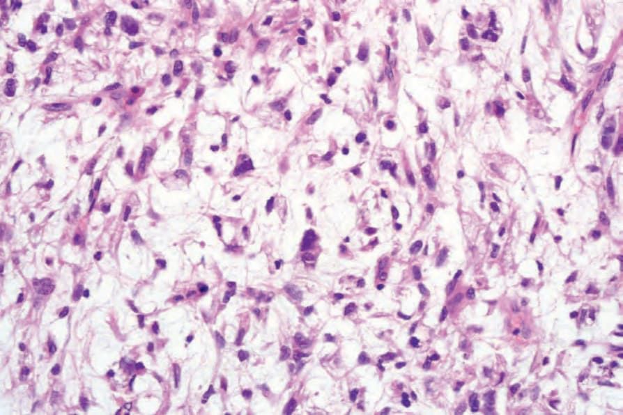
Fig. 35.195 Myxofibrosarcoma: note the pleomorphic tumor cells scattered in the myxoid matrix.
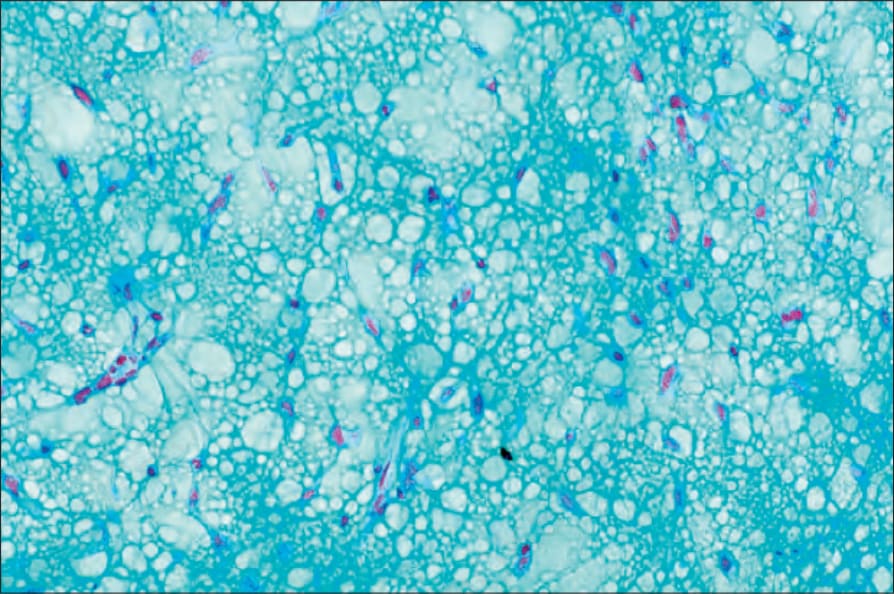
Fig. 35.196 Myxofibrosarcoma: the tumor stains strongly with Alcian blue at pH 2.5, indicating the presence of hyaluronic acid.
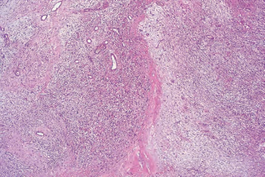
Fig. 35.197 Myxofibrosarcoma: in this field, the multinodularity is emphasized.
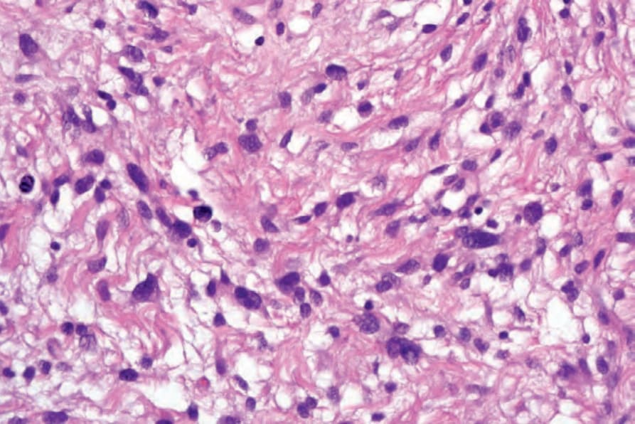
Fig. 35.198 Myxofibrosarcoma: there is marked nuclear pleomorphism.