Low-grade fibromyxoid sarcoma
Low-grade fibromyxoid sarcoma
Clinical features Low-grade fibromyxoid sarcoma is a rare distinctive tumor which belongs within the spectrum of hyalinizing spindle cell tumor with giant rosettes.1–20
Pathogenesis and histologic features The vast majority of cases are associated with t(7;16)(q33;p11) fusing CREB3L2 and FUS; the former can be substituted by t(11;16)(p11;p11) with FUS-CREB3L1 fusion.CREB3L1 (11p11) is seen in a small subset of cases.5,27–37 In some examples the fusion results in a supernumerary ring and these tumors appear to recur more frequently.27
Histology shows an infiltrative tumor with characteristic alternating myxoid and collagenous areas (Figs 35.188 and 35.189). Cellularity is
1755 Malignant fibroblastic tumors
not prominent and tumor cells tend to predominate in the myxoid areas. Bundles of bland, elongated spindle-shaped cells with focal whorling are seen. Small blood vessels with surrounding fibrosis are often present and tumor cells may concentrate around vascular channels. Cytologic atypia is minimal and mitotic figures are very rare. In some cases, there is transition to areas with tumor cells that are focally epithelioid surrounding prominently hyalinized collagen with formation of giant rosettes (Figs 35.190 and 35.191). The latter neoplasm was previously considered a distinct entity termed hyalinizing spindle cell tumor with giant rosettes.8,9,36 Rare cases contain focal areas resembling an ordinary fibrosarcoma or sclerosing epithelioid fibrosarcoma; it is still debated if the latter is related to low-grade fibromyxoid sarcoma (see also above).4–6,34,35,37 Recurrences may exhibit nuclear pleomorphism.21 A pediatric case mimicking ossifying fibromyxoid tumor has been documented.38
Differential diagnosis Distinction from myxofibrosarcoma is based on the presence of curvilinear blood vessels and at least focal prominent cytologic atypia with mitotic activity in the latter tumor.42 Tumor cells in neurofibroma are wavier and myxoid, and collagenous areas do not alternate, but focal collagen deposition is seen between tumor cells. Furthermore, tumor cells in neurofibroma are S100 protein positive. In addition, none of the benign or malignant mimickers, with the exception of sclerosing epithelioid fibrosarcoma, is positive for MUC4.
Immunohistochemistry shows staining for vimentin and very focal positivity for SMA and EMA. Claudin 1 is also often positive and this, coupled with positivity for EMA, may lead to a misdiagnosis of perineurioma.39 However, Glut-1 tends to be positive in perineurioma and negative in low-grade fibromyxoid sarcoma.40 MUC4 is a highly sensitive and specific marker of low-grade fibromyxoid sarcoma.41 Ultrastructural studies show cells with features of fibroblasts.
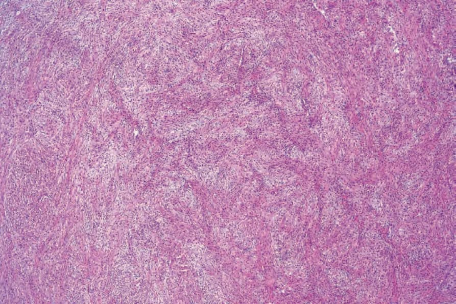
Fig. 35.188 Low-grade fibromyxoid sarcoma: this field shows cellular foci and adjacent myxoid regions.
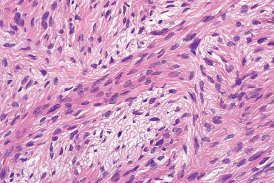
Fig. 35.189 Low-grade fibromyxoid sarcoma: high-power view.
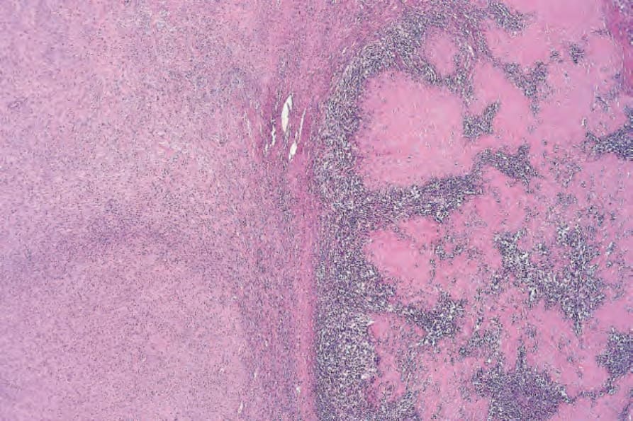
Fig. 35.190 Hyalinizing spindle cell tumor with giant rosettes: note the presence of giant rosettes containing abundant hyalinized collagen in the center.
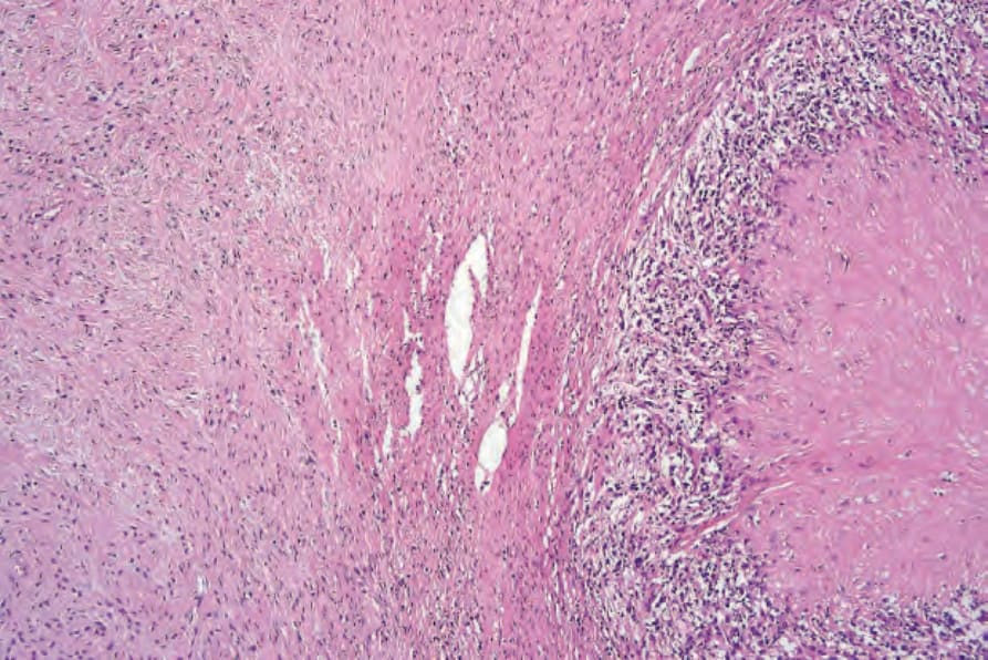
Fig. 35.191 Hyalinizing spindle cell tumor with giant rosettes: transition between bundles of bland elongated spindled cells and a giant rosette.