Sclerosing epithelioid fibrosarcoma
Sclerosing epithelioid fibrosarcoma
Clinical features Sclerosing epithelioid fibrosarcoma is a very rare distinctive variant of fibrosarcoma. It involves deep soft tissues of the lower limbs/limb girdles followed by the trunk and upper limbs and rarely the head and neck (including the salivary gland and mouth) and bone.1–6 Rare tumors can occur in the liver, colon, kidney, intra-abdominal soft tissues, pituitary gland, and skull.7–15 One tumor developed after radiotherapy.7 Patients are middle-aged adults and present with a variably painful mass. There is no sex predilection. There is a high recurrence rate of up to 50% and metastatic disease occurs in up to 40% of cases.1–6
Pathogenesis and histologic features Recently, this tumor has been described in combination with low-grade fibromyxoid sarcoma (see below) with demonstration of the characteristic t(7;16) (q33;p11) in these mixed cases.16,17 However, FUS rearrangements are uncommon in pure epithelioid fibrosarcoma, casting some doubt on the pathogenetic relationship between these two tumors in their pure, unmixed forms.18 Recent reports suggest that EWSR1-CREB3L1 rearrangements are predominant over FUS and CREB3L2 rearrangements in pure sclerosing epithelioid fibrosarcoma.19 In contrast, hybrid lesions recapitulate the genotype of low grade fibromyxoid sarcoma (most commonly, FUS-CREB3L2 fusion).19,20
The tumor is characterized by prominent hyalinization and relatively uniform, small, round or ovoid epithelioid cells with sparse and often clear cytoplasm arranged in cords and nests (Figs 35.186 and 35.187). Areas of typical fibrosarcoma may be seen. Focal calcification and bone formation are features that are sometimes present.
It presents mainly in young adults or less commonly younger patients, including children as a slowly growing, asymptomatic large mass with a predilection for the limbs.1–7,20,21 Most lesions are deep seated, may be intramuscular and some tumors are subcutaneous. Superficial lesions tend to be more common in children.10 Unusual sites include intracranial, intrathoracic, mesenteric, omental, the ovary, the lung, the colon, the external anal sphincter and the falciform ligament.11,22–24 Although the rates of local recurrence, metastasis and death were high in the first published series, a subsequent large series reported the rates of local recurrence, metastasis and death as 9%, 6%, and 2%, respectively.3,4,6 Local recurrence and metastases may occur many years after excision of the primary tumor.1,3,6,19–21 Cases associated with radiotherapy have been reported.25,26
Tumor cells are generally positive for vimentin and beta-catenin. MUC4 is positive in the majority of cases; MUC4-positive cases are reported to be associated with a FUS gene rearrangement.21 Some show focal positivity for EMA, S100 protein and more infrequently for neuron-specific enolase (NSE).1,3 Rare tumors focally express keratin.1
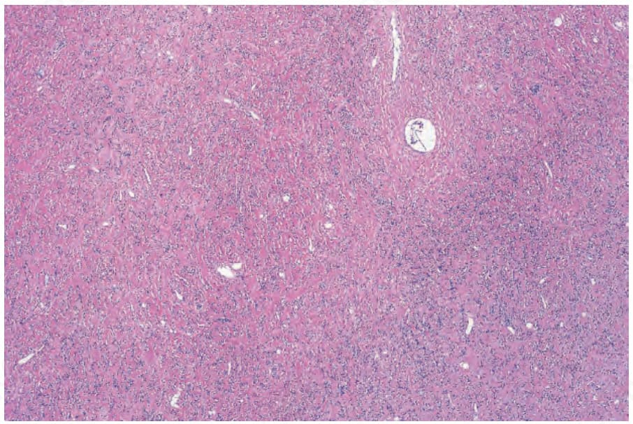
Fig. 35.186 Sclerosing epithelioid fibrosarcoma: low-power view showing a paucicellular infiltrate within a densely hyalinized stroma.
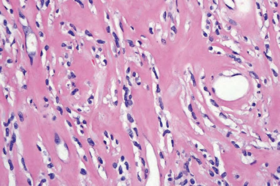
Fig. 35.187 Sclerosing epithelioid fibrosarcoma: the tumor cells characteristically show a single-file distribution.