Fibrosarcoma: adult variant
Fibrosarcoma: adult variant
Clinical features Contrary to age-old teaching, adult fibrosarcoma is now regarded as distinctly uncommon. With the aid of improved diagnostic techniques, many previous cases of fibrosarcoma would now be reclassified, most often as monophasic synovial sarcoma, solitary fibrous tumor or malignant peripheral nerve sheath tumor. True fibrosarcoma accounted for less than 1% of sarcomas among 100 000 adult sarcoma patients studied.1
Adult fibrosarcoma usually arises in the fifth and sixth decades, and shows a slight male predominance. It occurs most often in the lower limbs, followed by the upper limbs and trunk.1–4 Tumors presenting in children are classified separately (see below). Fibrosarcoma is most often deep seated and asymptomatic. Only very occasional tumors are subcutaneous. There is a tendency for local recurrence and metastasis, with a 5-year survival of about 50%.
fibrillary xanthomatous or glassy cytoplasm; intranuclear and cytoplasmic inclusions are sometimes found. Focal granular cell change is frequent (Fig. 35.183). Arborizing vessels and a mixed inflammatory infiltrate are common features.1,2
Pathogenesis and histologic features A small proportion of adult tumors have been reported to be radiation induced but it is not clear whether all of these tumors represent true fibrosarcomas.5
Cytogenetic studies in a small number of fibrosarcomas have shown complex chromosomal abnormalities.6,7 In two cases, tri- or tetrasomy of 2q has been described.6
Adult fibrosarcoma tends to be well circumscribed and is composed of relatively uniform spindled cells with little cytoplasm, typically arranged in a herring-bone pattern (Figs 35.184 and 35.185). There is usually minimal collagen production, mild pleomorphism can be present and the mitotic count varies. It is important to remember that tumors such as dermatofibrosarcoma protuberans and dedifferentiated liposarcoma may have areas identical to fibrosarcoma.
Immunohistochemically, tumor cells are diffusely positive for CD34; focal staining for cytokeratin is seen in most cases.1,2 FLI-1, ERG, S100 protein, desmin, and smooth muscle actin are negative. There is no overexpression of TP53 and SMARCB1 is retained.
Differential diagnosis The combination of pleomorphic histology with very low mitotic index and diffuse CD43 expression in a superficial location is fairly distinctive. The main differential diagnosis is with myxoinflammatory fibroblastic sarcoma. In myxoinflammatory fibroblastic sarcoma, inflammatory cells are prominent, a number of cells display virocyte-like nuclear inclusions, and expression of CD34 is more focal. Cytogenetics studies may be of help in difficult cases (see also myxoinflammatory fibroblastic sarcoma).
Immunohistochemistry shows that tumor cells are positive for vimentin and are occasionally focally positive for actin. Other markers including CD34, S100 protein, EMA and desmin are negative.
Ultrastructural studies show cells with features of fibroblasts and myofibroblasts.8
Differential diagnosis All cases should be assessed for S100 protein, pankeratin and EMA expression to exclude malignant schwannoma and monophasic synovial sarcoma since the diagnosis of fibrosarcoma is one of exclusion. Leiomyosarcoma is composed of plumper spindle-shaped cells with abundant eosinophilic cytoplasm
1754 Connective tissue tumors
and cigar-shaped nuclei. Tumor cells are usually positive for desmin and staining for actin is more widespread than that seen in fibrosarcoma.
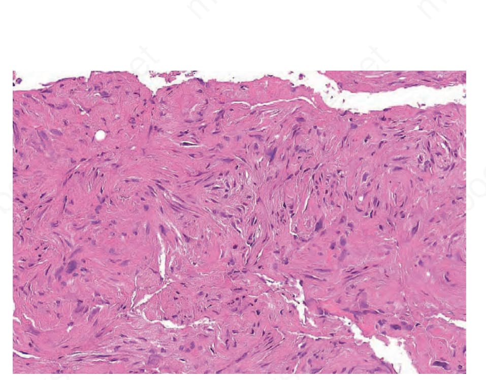
Fig. 35.183 Superficial CD34-positive fibroblastic tumor: extensive replacement of the subcutaneous tissue by a predominantly spindle cell tumor.
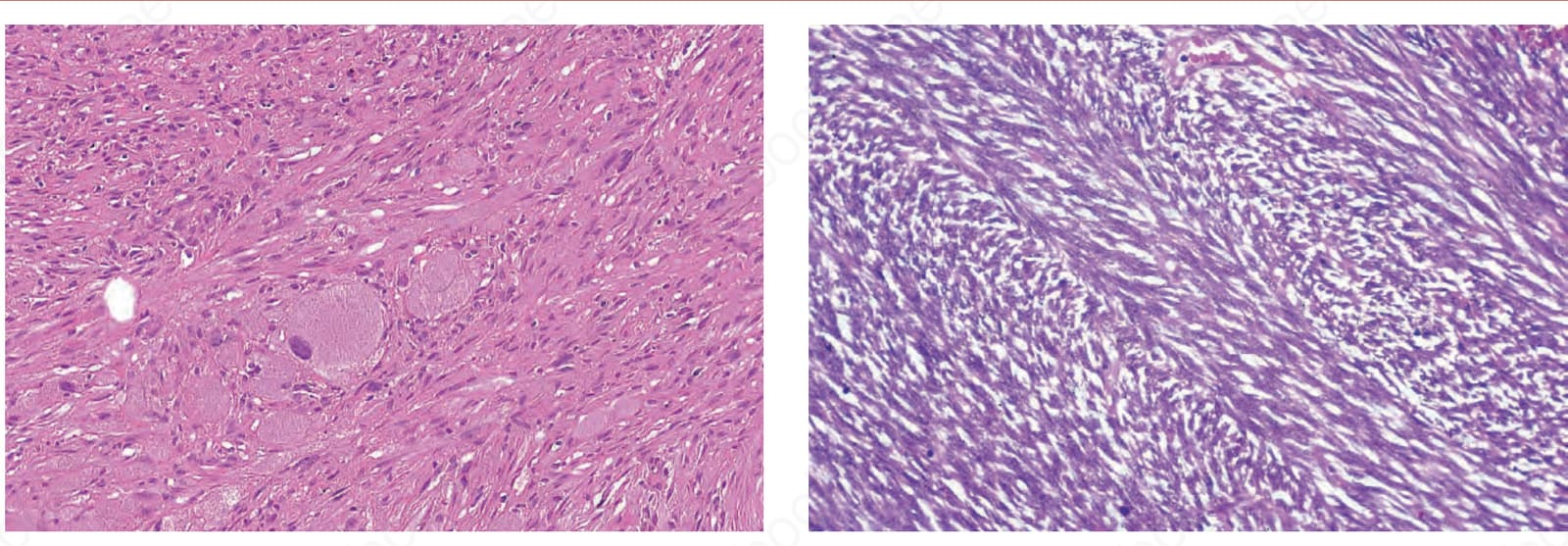
Fig. 35.184 Fibrosarcoma: marked basophilia and a herring-bone pattern of spindle cells are typical features of this lesion.
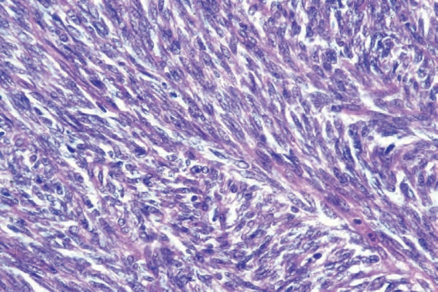
Fig. 35.185 Fibrosarcoma: the spindle cell borders are indistinct and their nuclei are elongated with thin tapered ends, unlike those in leiomyosarcoma and neurofibrosarcoma.
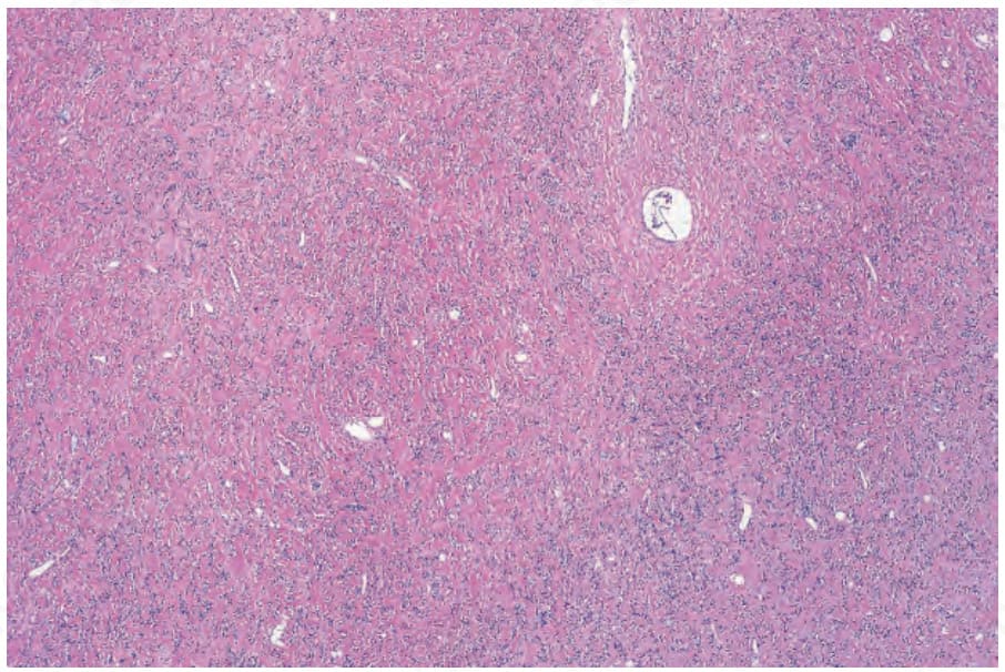
Fig. 35.186 Sclerosing epithelioid fibrosarcoma: low-power view showing a paucicellular infiltrate within a densely hyalinized stroma.