Giant cell angiofibroma
Giant cell angiofibroma
Clinical features Giant cell angiofibroma is a distinctive form of a giant cell-rich variant of solitary fibrous tumor occurring mainly in the orbit but also presenting elsewhere in the head and neck (including the oral cavity and pharynx), trunk, groin, vulva and perianal area.1–9 An exceptional case in the mediastinum has also been described.8 Patients are usually middle-aged adults and although orbital lesions have predilection for males, extraorbital lesions affect mainly females.1–5
1751 Intermediate (rarely metastasizing) fibroblastic and myofibroblastic tumors
slight male predilection.1–4 Tumors may rarely occur in bone, the oral cavity, heart, abdomen, stomach, breast, vulva, thoracic spine and larynx.1–3,5–25 A single cutaneous tumor has been described.12 A case in association with a desmoplastic melanoma has been reported.26 Multicentricity is exceptional.27 Local recurrence is common and metastatic disease is rare.1,23,24
Histologic features Histology shows an infiltrative tumor composed of fascicles of cells with indistinct cytoplasmic margins, pale pink cytoplasm and elongated, vesicular nuclei. In some lesions there is variable cellularity. Cytologic atypia is not prominent but is always present. Mitotic activity tends to be low. In a single case intracytoplasmic hyaline inclusions have been reported.28 It has been suggested that in tumors of the head and neck, aggressive behavior is associated with higher mitotic activity and necrosis.29
Tumors are asymptomatic, subcutaneous, slowly growing and measure only a few centimeters in diameter. Local recurrence is rare. Aggressive behavior, however, has not been documented in any cases.
Pathogenesis and histologic features Cytogenetic studies in a one case showed abnormalities of chromosome 6q, while another showed t(12;17)(q15;q23).10,11 NAB2-STAT6 fusion transcripts have been identified in some cases of solitary fibrous tumor with giant cell angiofibroma-like features.12
Tumor cells are positive for smooth muscle actin, muscle specific actin and calponin.30,31 There may be focal positivity for desmin but h-caldesmon is negative.30 ALK-1 and keratin are also negative a feature that allows distinction from inflammatory myofibroblastic tumor.30 Beta-catenin shows no nuclear staining, a feature that allows distinction from desmoid fibromatosis.
Ultrastructural examination reveals subplasmalemmal bundles of myofilaments and fibronexus.32
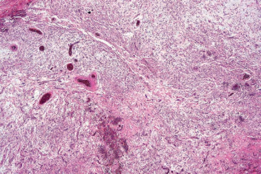
Fig. 35.175 Giant cell angiofibroma: low-power view showing prominent blood vessels and tumor cells dispersed in a myxoid stroma.
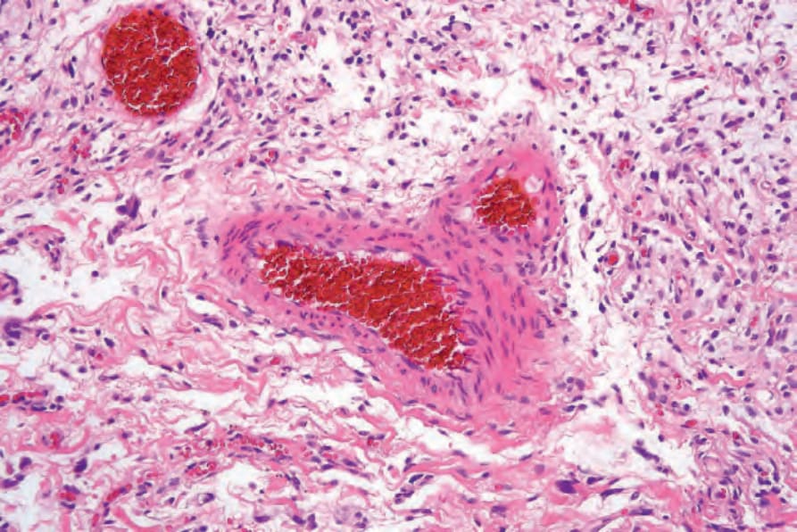
Fig. 35.176 Giant cell angiofibroma: high-power view.
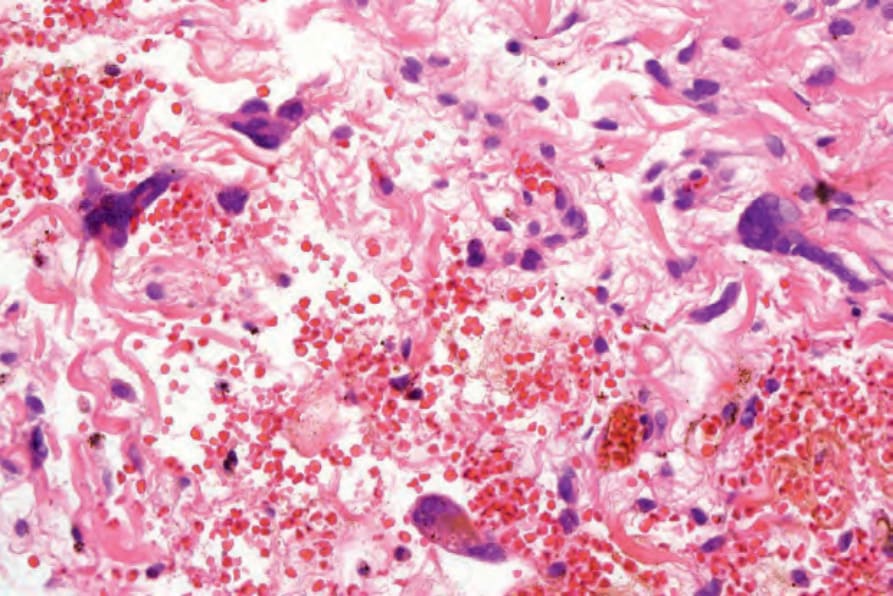
Fig. 35.177 Giant cell angiofibroma: note the tumor giant cells.