Plantar fibromatosis
Plantar fibromatosis
Clinical features Plantar fibromatosis (Ledderhose disease) is essentially the equivalent of palmar fibromatosis affecting the foot (although flexion deformity only rarely develops). The condition may be bilateral, usually arising asynchronously.1,2 Although the overall age and sex distribution is similar to palmar fibromatosis, a significant proportion of cases occur in children or adolescents.3 Familial cases including occurrence in twins are rare.4,5 It is much less common than Dupuytren contracture and may be associated with it and with diabetes.6 Characteristically, it presents as single or multiple nodules on the medial aspect of the sole, usually just distal to the pedal arch. A variant of plantar fibromatosis presenting in children and characterized by nodules on the anteromedial aspect of the sole has been documented.7 Involvement of the plantar aspect of the heel in children is also seen.8
A heterozygous missense variant in the Asteroid Homolog 1 (ASTE1) has been described in a family with LMNA-related cardiomyopathy and palmar and plantar fibromatosis.32
The microscopic appearances of palmar fibromatosis depend, to some extent, upon the duration of the lesion. In the early stages, cellular nodules composed of uniformly plump, proliferating myofibroblasts develop in the palmar aponeurosis, and may show mitotic activity but without atypia (Figs 35.126 and 35.127). This proliferative process, which has very little collagenous stroma, gradually extends as an infiltrative mass into adjacent subcutaneous tissues.
Although most cases are asymptomatic, patients may complain of discomfort or a burning sensation, particularly after walking. Neurological symptoms due to entrapment of nerves are exceptionally encountered.9 Contracture of the toes is extremely rare.10 Local recurrence is very common.3,11 Plantar hyperkeratosis is a rare association.12
1738 Connective tissue tumors
Pathogenesis and histologic features Cytogenetic studies have revealed trisomy 8 and trisomy 14.13 Superficial fibromatoses are genetically distinct from deep fibromatoses in their lack of mutations in CTNNB1, the gene encoding β-catenin.14–16
The histologic and features are very similar to those of palmar fibromatosis; however, evidence of chronic inflammation or previous hemorrhage (both of which are probably secondary in nature) is more frequently present in plantar fibromatosis. Lesions also tend to be more consistently cellular and show much less tendency to hyalinize with time (Fig. 35.129). Scattered multinucleated giant cells may be seen.17 Osseous metaplasia is exceptional.18
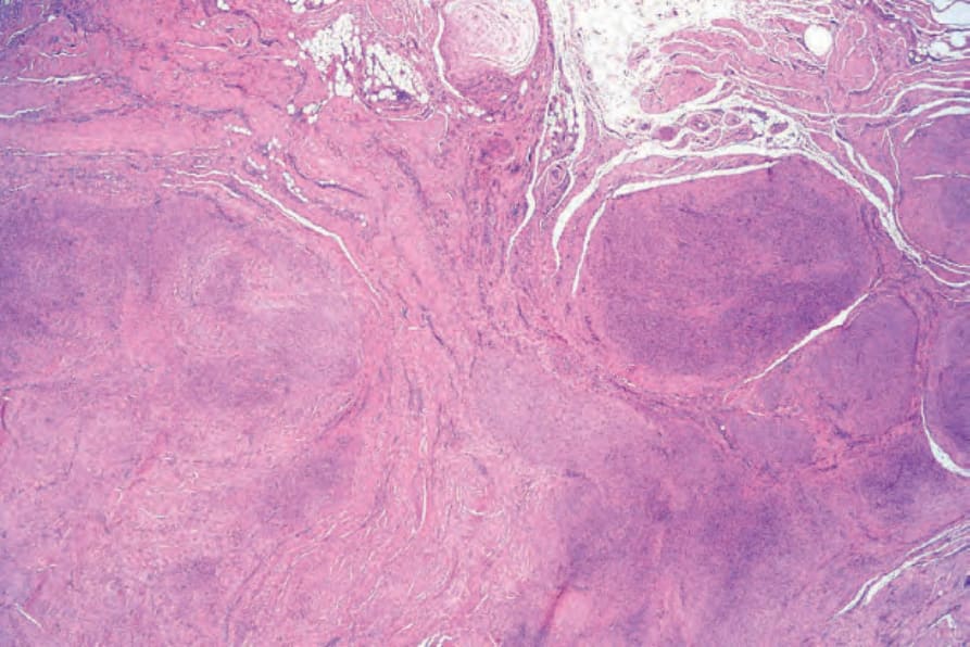
Fig. 35.126 Palmar fibromatosis: scanning view showing multiple nodules of hypercellular tissue with admixed hyalinized collagen.
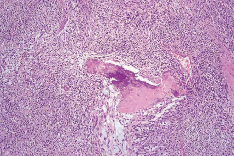
Fig. 35.129 Plantar fibromatosis: this example is strikingly cellular and shows focal osseous metaplasia.

Fig. 35.130 Penile fibromatosis: biopsy from end-stage disease showing dense collagen without any significant inflammation. Despite its name, the condition most probably represents a reactive process.