Juvenile hyaline fibromatosis
Juvenile hyaline fibromatosis
Clinical features Juvenile hyaline fibromatosis is an exceedingly rare autosomal recessive disfiguring condition of younger children and usually presents as cutaneous papules and nodules, multiple soft tissue masses of variable size that particularly affect the head and neck, and gingival hyperplasia (Fig. 35.122).1–7 The back and flexures of the lower limbs may also be involved, resulting in flexion contractures. Often, joint contractures and gingival hypertrophy precede the cutaneous manifestations of the disease.8 Other associations include mental retardation and osteolytic bone lesions, hyperpigmentation, osteosclerosis, scoliosis, atrial thrombus, and pericardial effusion.9–12 A more localized and limited form of the disease has been documented.13,14 In a single case, a squamous cell carcinoma developed in association with oral lesions.15
and lower limb.1–6 About 10% of cases occur in an intra-abdominal/retroperitoneal location.2 There seems to be a predilection for tumors to occur along the milk line. Simple excision is the treatment of choice and there is no tendency for local recurrence.2
The only treatment is surgical excision of each lesion, but new tumors may continue to develop into adult life. Targeted therapy with proteasome inhibitors has been suggested as a therapeutic option.16
Pathogenesis and histologic features Cytogenetic analysis has shown deletion or rearrangement of 13q14 that translates into loss of RB1 expression. Mammary-type myofibroblastoma appears to be part of a morphological spectrum that includes cellular angiofibroma and spindle cell lipoma (as well as pleomorphic lipoma and pleomorphic fibroma). These former tumors not only share morphological features but also the same cytogenetic abnormality.2,7,8 There does not seem to be a relationship between solitary fibrous tumor and mammary-type myofibroblastoma.9
Tumors are well-circumscribed and composed of a mixture of bland spindled cells and variable, sometimes prominent, amounts of mature adipose tissue.1 The spindled cells are arranged in fascicles and display a myofibroblast-like appearance with tapering nuclei and amphophilic cytoplasm with an indistinct cytoplasmic membrane. Nuclear palisading as seen
Infantile systemic hyalinosis is considered to be an allelic disorder with similar but more severe involvement, hyaline deposits in many organs, recurrent infections and death early in life, usually within the first 2 years.17–21
Pathogenesis and histologic features Juvenile hyaline fibromatosis, thought to be due to a genetic abnormality of collagen production, may be seen in siblings, particularly of consanguineous parents.2,22–24 Ultrastructural studies and skin fibroblasts from cultures have suggested defective synthesis of collagen within fibroblasts.25–27 The disease results from an abnormal assembly of basement membrane material and collagen deposition is also abnormal.19,20,28 The gene for the disease has been mapped to chromosome 4q21.29 Multiple different mutations have been identified in this gene, which encodes capillary morphogenesis protein 2 (CMG2), also termed anthrax toxin receptor 2 (ANTXR2).30–35 ANTXR2 is a transmembrane protein induced during capillary morphogenesis.19,20 The
1736 Connective tissue tumors
myofibromatosis presents with multiple lesions but the histologic features are quite different from those of juvenile hyaline fibromatosis. Nuchal fibroma presents as a single lesion and is characterized by abundant collagen lacking masses of amorphous eosinophilic material.
Intermediate (locally aggressive) fibroblastic and myofibroblastic tumors
Locally aggressive fibrous lesions are defined as infiltrative neoplasms that are prone to local recurrence. They may be destructive, but never metastasize.
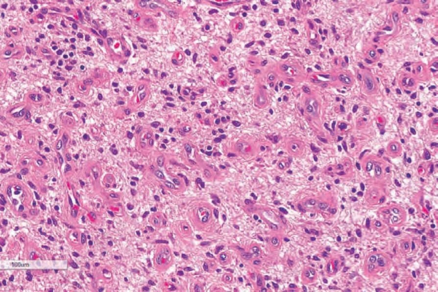
Fig. 35.121 Angiofibroma of soft tissue: abundant myxoid stroma. By courtesy of C.D.M. Fletcher, MD, Brigham and Women’s Hospital and Harvard Medical School, Boston, USA.
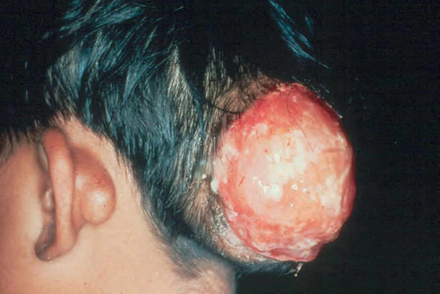
Fig. 35.122 Juvenile hyaline fibromatosis: there is a large circumscribed ulcerating mass. A smaller lesion is also present behind the left ear. By courtesy of Y. Kitano, MD, Osaka University School of Medicine, Japan.
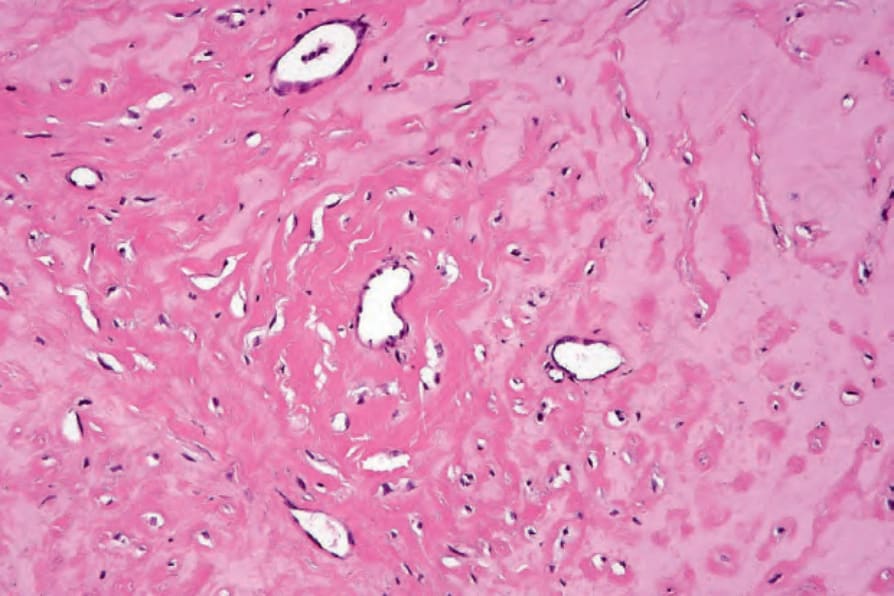
Fig. 35.123 Juvenile hyaline fibromatosis: this field is largely acellular and composed of intensely eosinophilic, hyalinized material. By courtesy of J. Sciubba, DDS, Baltimore, USA.
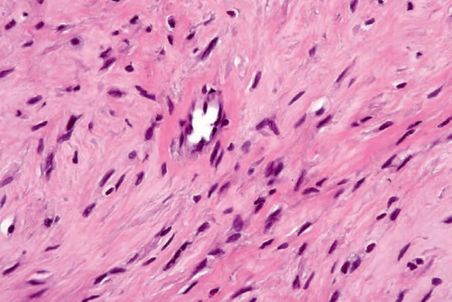
Fig. 35.125 Juvenile hyaline fibromatosis: the spindle cells are bland with hyperchromatic nuclei. By courtesy of J. Sciubba, DDS, Baltimore, USA.