Acquired digital fibrokeratoma
Acquired digital fibrokeratoma
Clinical features Acquired digital fibrokeratoma is of no clinical significance and arises most often in adult life, affecting males more often than females.1–3 It presents as a slowly growing firm nodule or excrescence, usually less than 1 cm in size, on the fingers or toes; as such, it may clinically resemble a supernumerary digit (Fig. 35.111). Periungual lesions are known as acquired periungual fibrokeratomas. A case with multiple lesions has been reported.4 A similar lesion has been documented on the heel.5,6 Some regard these lesions as traumatic, although with little supporting evidence. A case associated with ciclosporine treatment has been documented.7 Acquired digital fibrokeratoma may be similar to the periungual fibroma that occurs in tuberous sclerosis but the latter tends to be multiple and has minimal or no epidermal component.8
Histologic features Tumors are characterized by a band-like proliferation of fibroblast-like bland cells usually occupying the upper part of the dermis. Extension into the subcutaneous tissue is rare. Scattered small dilated vascular channels are present and tumor cells are interspersed with collagen bundles with decrease in elastic fibers. Superficial tumor cells have a vertical orientation to the epidermis and deeper cells usually have a horizontal orientation with regards to the epidermis. CD34 is positive in tumor cells and there is minimal focal staining for smooth-muscle actin and few factor XIIIa-positive cells.
Histologic features Microscopically, fibrokeratomas are pedunculated lesions covered by variably acanthotic and hyperkeratotic skin (Figs 35.112 and 35.113). The core is composed of an admixture of dense collagen fibers containing a variable
Differential diagnosis The main differential diagnosis is with early plaque-stage dermatofibrosarcoma protuberans. This is discussed under the latter condition.
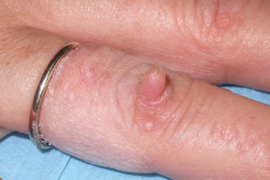
Fig. 35.111 Acquired digital fibrokeratoma: the resemblance to a supernumerary digit is striking. By courtesy of J.C. Pascual, MD, Alicante, Spain.
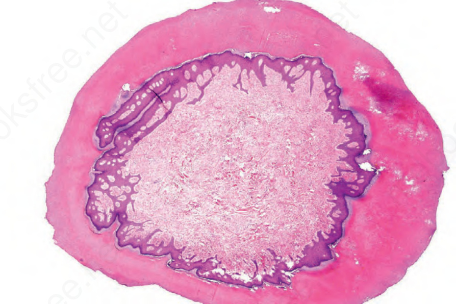
Fig. 35.112 Acquired digital fibrokeratoma: low-power view showing the hyperkeratotic acanthotic surface epithelium.