Liposarcoma
Liposarcoma
Clinical features Despite being one of the most common soft tissue sarcomas, liposarcoma presents infrequently as a primary subcutaneous lesion and only exceptionally as a primary dermal tumor.1
Liposarcoma is traditionally divided into three subtypes:
• well-differentiated / dedifferentiated 2–7
• myxoid, which includes the cellular (previously termed round cell) variant,2–5,8,9
• pleomorphic.2–5,8–10
Dedifferentiated liposarcoma Dedifferentiation refers to a biphasic tumor containing an atypical lipomatous tumor and a sarcomatous undifferentiated component of variable grade. Dedifferentiation occurs mainly in deep-seated tumors (particularly those occurring in the retroperitoneum) and is exceptional in subcutaneous lesions.11,12
Myxoid liposarcoma Myxoid liposarcoma occurs in adults, with a peak incidence between the fourth and fifth decades of life, and has no sex predilection.9,13,14 Although rare in children and adolescents, it is the most common type of liposarcoma in this age group.15 The majority of tumors arise in deep soft tissues, subcutaneous tumors being very rare.16 The most common site is the lower limb, particularly the thigh. All tumors, without exception, tend to local recurrence, and metastases occur in approximately one-third of cases depending on the grade.13,14,17 The presence of a cellular (previously termed round cell) component, necrosis and p53 overexpression has been found to be associated with poor prognosis.17 The terminology of round cell liposarcoma is no longer favored under the WHO classification as round cell is not the only morphology adopted by the cellular (higher grade) variant of myxoid liposarcoma.
Pleomorphic liposarcoma Pleomorphic liposarcoma is the least common variant of liposarcoma. There is no sex predilection, patients are elderly and tumors are deep seated, presenting mainly on the limbs.18,19 Only exceptional tumors occur in the dermis or subcutis.1,10,18,19 Tumors grow rapidly and there is a high tendency for local recurrence and metastasis.18,19 While being by a wide margin the least common liposarcoma in deep soft tissue, it may be the most common liposarcoma to occur as a cutaneous primary.10
Pathogenesis and histologic features Dedifferentiated liposarcoma shares the same cytogenetic alterations with atypical lipomatous tumor (well-differentiated liposarcoma), namely a ring or giant marker chromosome associated with amplification and overexpression of the 12q13~15 region containing MDM2. In addition, JUN (1p32), ASK1 (6q23) and TAB2 (6q25) amplification can be seen..20–24
Myxoid/round cell liposarcomas show a specific t(12;16)(q13;p11) fusing DDIT3 and FUS. In a small subset of cases a t(12;22)(q13;q12) has been found where EWSR1 substitutes for FUS. These translocations are seen in virtually all cases.20–22,24–28
No consistent cytogenetic abnormality has been demonstrated in pleomorphic liposarcoma, which often shows complex cytogenetic abnormalities with TP53 mutations.23,24,27
Dedifferentiated liposarcoma is defined as a well-differentiated liposarcoma showing abrupt transition to a higher grade nonlipogenic sarcoma.29
1713 Malignant adipocytic tumors
Less frequently the transition may be to a non-lipogenic low-grade sarcoma, but this terminology and the behavioral features are debated. In some cases, a well-differentiated area cannot be identified. Focal lipoblastic differentiation may be seen within the dedifferentiated areas.30 This change can occur in a primary tumor and less often in a recurrence. Although in most cases the dedifferentiated component is pleomorphic, there are often focal less atypical areas that may mimic other tumors such as dermatofibrosarcoma protuberans. Moreover, local recurrences of dedifferentiated liposarcoma may be well-differentiated. Meningothelial-like whorls have also been documented.31,32 Heterologous differentiation including osteosarcomatous, chondrosarcomatous, myogenic, and angiosarcomatous components is rarely seen.33 Positive diffuse staining of MDM2 and/or CDK4 is a constant finding.
extramedullary hematopoiesis may be seen. The presence of more cellular areas composed of uniform oval-to-round larger cells with hyperchromatic nuclei and inconspicuous cytoplasm indicates round cell change. This is associated with more aggressive behavior and such lesions are known either as combined myxoid and round cell liposarcoma or as high-grade myxoid liposarcoma (Figs 35.46 and 35.47).5,6,29 The round cell component may predominate. Pure round cell liposarcomas are extremely rare in the subcutis, but mixed tumors are occasionally seen. Dedifferentiation in myxoid liposarcoma is exceptional.34 S100 is positive in lipoblasts and variably positive in the round cell component. Immunohistochemistry for the cancer testis antigen NY-ESO-1has been reported to be consistently positive in this group of tumors but not in other lesions that may be confused with them.35
Myxoid liposarcoma is composed of fairly uniform stellate or spindled cells with small vacuoles set in a myxoid matrix composed of acid mucopolysaccharide (Figs 35.42–35.45).3,5,13,14 Mucin pooling, producing a lymphangioma-like pattern, is common. Emphasis should be placed on the presence of a complex plexiform network of small thin-walled capillaries in a pattern resembling chicken-wire or ‘crow’s feet.’ Mitoses are sparse. Lipoblasts are most easily identified at the periphery of the tumor. Occasionally,
Pleomorphic liposarcoma consists of highly pleomorphic spindle cells, lipoblasts, and numerous multinucleated multivacuolated giant cells (Fig. 35.48).18,19,36 Identification of lipoblasts is vital in distinguishing this variant from other pleomorphic sarcomas.
Lipoblasts are variable present within all of these lesions and can be absent or very difficult to find in myxoid and well-differentiated liposarcoma. By definition, they are absent in the dedifferentiated liposarcoma component which is nonadipocytic. In exceptional cases, the dedifferentiated portion
1714 Connective tissue tumors
of liposarcoma can have a pleomorphic liposarcoma-like component, but 12q15 amplification and MDM2 and CDK4 expression are maintained. In pleomorphic liposarcoma proper, lipoblasts can range from diffusely present to very rare and focal. Typically, lipoblasts are highly variable in size, contain more than one well-defined or punched-out lipid vacuole, and have irregular, hyperchromatic (and sometimes multiple) nuclei, the margins of which are scalloped by the fat droplets.
Differential diagnosis Nuclear positivity for MDM2 and or CDK4 will help establish the diagnosis of dedifferentiated liposarcoma in most cases (see also atypical lipomatous tumor).37,38 Myxoid liposarcoma is distinguished from other sarcomas by its distinctive vessel morphology. In difficult cases molecular confirmation may be necessary.16 Myxofibrosarcoma (myxoid malignant fibrous histiocytoma) is recognized by the lack of lipoblasts and the presence of more variable pleomorphism than present in myxoid liposarcoma. Distinction from lipoblastoma may be very difficult or impossible. However, liposarcoma is very rare in children and, in difficult cases, cytogenetic studies may be helpful (see lipoblastoma). The diagnosis of pleomorphic liposarcoma is based on the identification of lipoblasts in the background of a pleomorphic sarcoma and a lack of MDM2 and/or CDK4 nuclear positivity.
TUMORS OF FIBROUS, FIBROBLASTIC AND MYOFIBROBLASTIC TISSUE
Benign fibrous and myofibroblastic tumors and tumorlike lesions
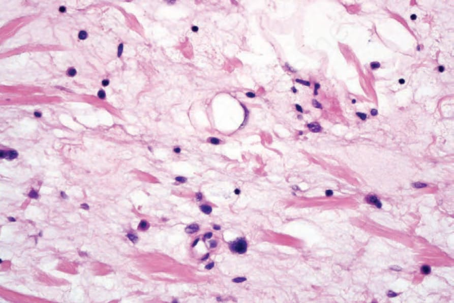
Fig. 35.40 Atypical spindle cell lipomatous tumor: high-power view showing signet-ring cell lipoblasts.
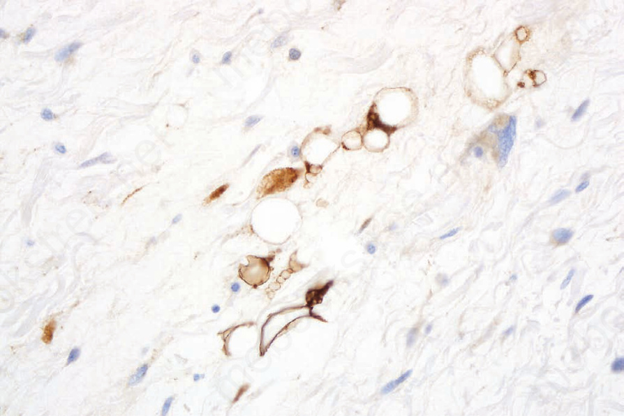
Fig. 35.41 Atypical spindle cell lipomatous tumor: the lipoblasts can be highlighted with S100 protein immunohistochemistry.
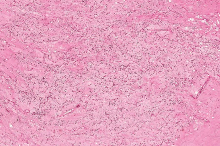
Fig. 35.42 Myxoid liposarcoma: low-power view showing spindled cells in a myxoid stroma and conspicuous delicate vessels.
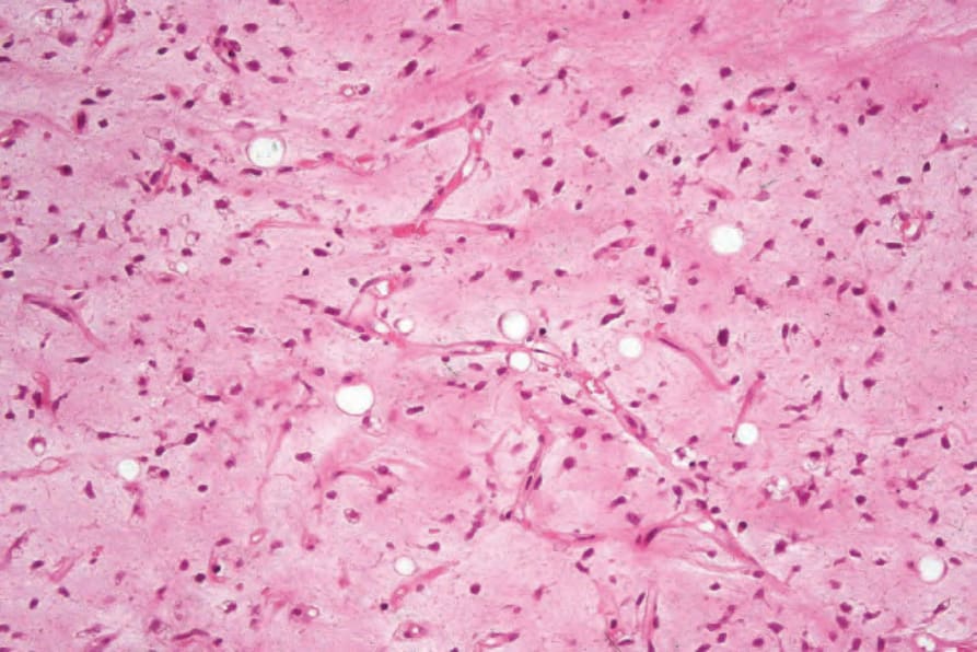
Fig. 35.43 Myxoid liposarcoma: medium-power view showing multiple signet-ring cell lipoblasts and conspicuous capillaries.
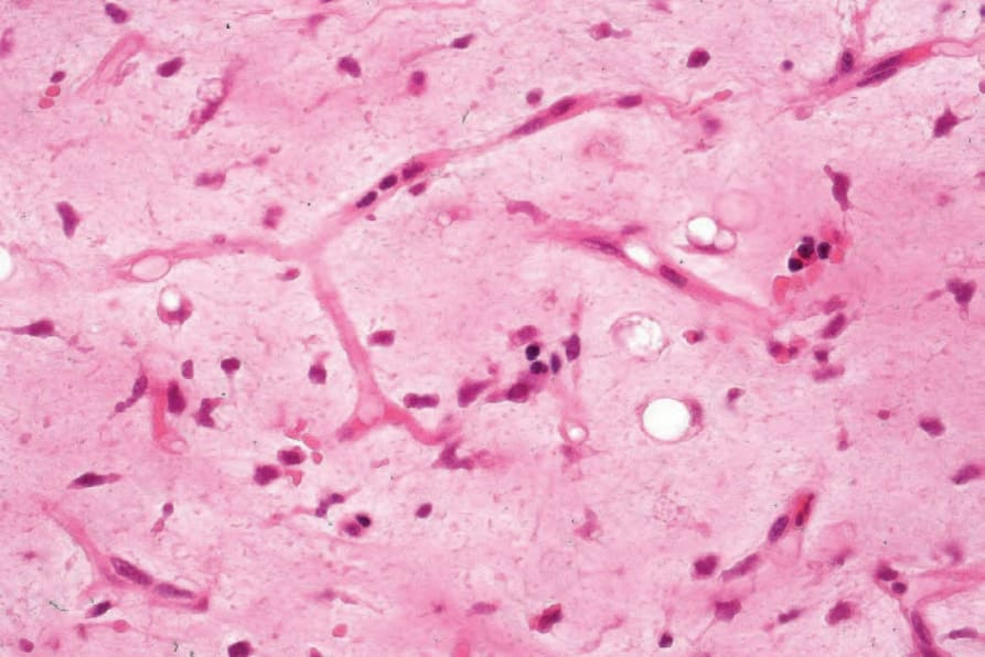
Fig. 35.44 Myxoid liposarcoma: this field shows a typical ‘crow’s-foot’ type of blood vessel.
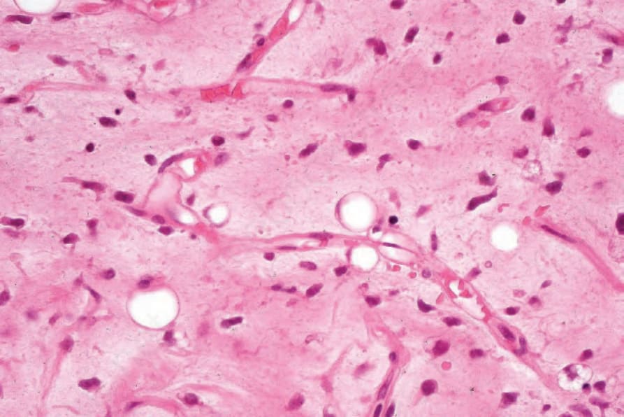
Fig. 35.45 Myxoid liposarcoma: multiple lipoblasts are present.
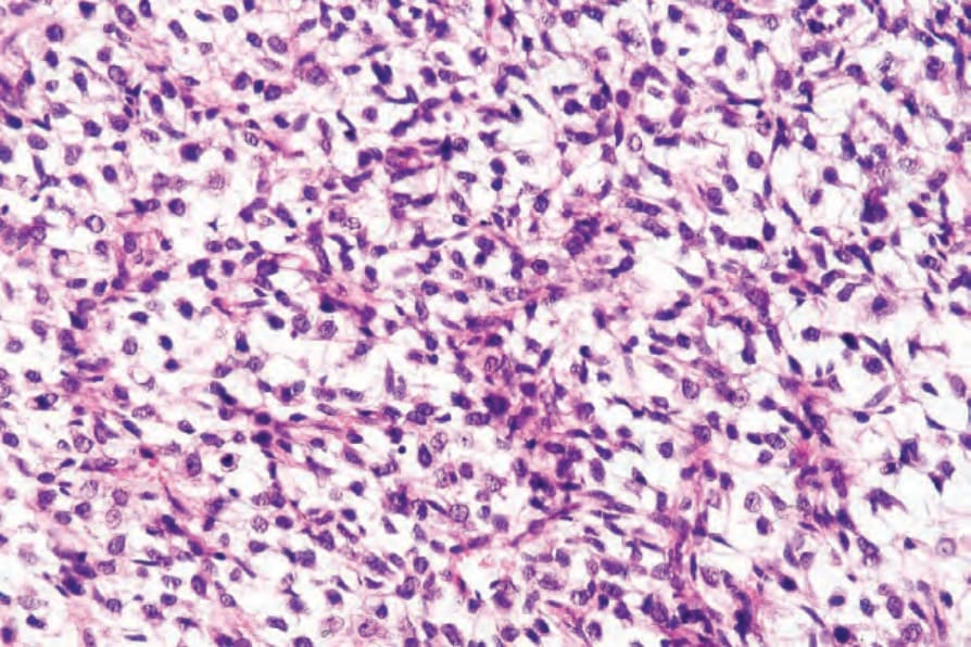
Fig. 35.46 Myxoid liposarcoma: round cell component. In this field, the tumor is much more cellular. The background vasculature is still visible.
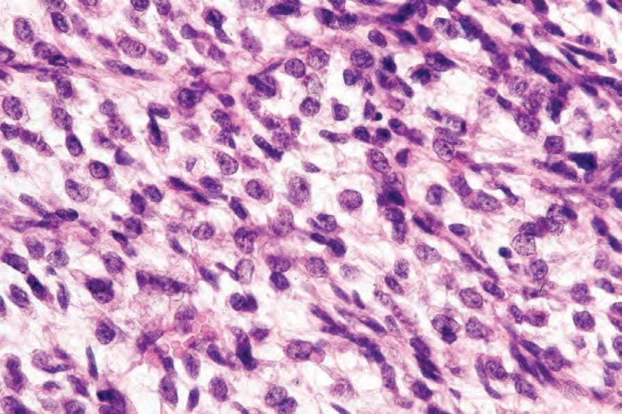
Fig. 35.47 Myxoid liposarcoma: round cell component. The nuclei are hyperchromatic and pleomorphic.
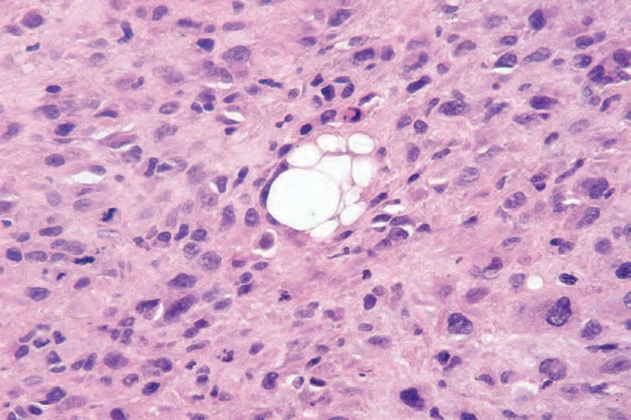
Fig. 35.48 Pleomorphic liposarcoma: recognition of this variant is dependent upon identification of lipoblasts among the highly pleomorphic cellular background.