Extraskeletal mesenchymal chondrosarcoma
Extraskeletal mesenchymal chondrosarcoma
Clinical features Extraskeletal mesenchymal chondrosarcoma is a very rare, deep-seated tumor that tends to occur more often in younger adults than the myxoid type.1–7 Children may be affected.8 It is more common in females and appears frequently in the head and neck region and upper trunk, and less often in the limbs. The prognosis appears to be worse than that for the myxoid variants.
Pathogenesis and histologic features In recent years, it has become clear that this tumor does not have chondrosarcomatous lineage as previously believed and it is more likely to represent a tumor of primitive mesenchymal cells.7,25
Extraskeletal myxoid chondrosarcoma is characterized in most cases by a specific t(9;22)(q22;q12) fusing NR4A3 and EWSR1.26–29 EWSR1 can be substituted by at least three additional homologous genes in a small subset of cases. Tumors with variant NR4A3 gene fusions are related with higher grade, rhabdoid phenotype, and a less favorable outcome compared with the EWSR1-NR4A3 positive tumors.30–45 A single case with HSPA8 as a fusion partner of NR4A3 has been recently reported.46 Loss of nuclear INI1 (encoded by SMARCB1) expression is documented in a subset of cases, particularly those with epithelioid morhology.47
These tumors have a characteristic microscopic appearance consisting of irregular but well-defined lobules with peripheral closely packed undifferentiated small cells with little cytoplasm (Figs 35.637 and 35.638). The
Pathogenesis and histologic features An inv(8) (q13q210) resulting in a recurrent HEY1-NCOA2 fusion gene has been identified.9
Extraskeletal mesenchymal chondrosarcoma has a distinctive biphasic histologic appearance characterized by undifferentiated round or spindle-shaped mesenchymal cells and variably prominent foci of generally mature, well-differentiated cartilage (Figs 35.640 and 35.641). Mitotic activity is usually prominent. Often, the undifferentiated cells are arranged around numerous slit-like vessels in a hemangiopericytoma-like pattern. Cartilaginous areas may show dystrophic calcification and sometimes ossification.
Tumor cells are positive for CD99 and Bcl-2.10 The cartilaginous areas are S100 protein positive. ERG positivity may also be seen.11 Nuclear Sox9, a chondrogenic transcription factor, can be detected.4,7,12–14
1878 Connective tissue tumors
MISCELLANEOUS REACTIVE, BENIGN LESIONS AND TUMORS OF UNCERTAIN DIFFERENTIATION
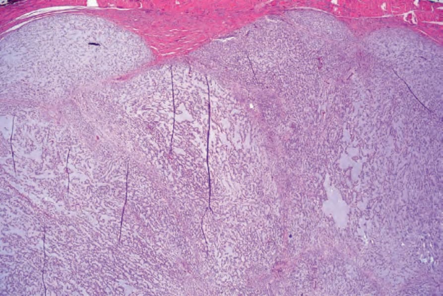
Fig. 35.637 Extraskeletal myxoid chondrosarcoma: the tumor is lobulated and shows a biphasic population. Small hyperchromatic cells at the periphery merge with a central myxoid component.
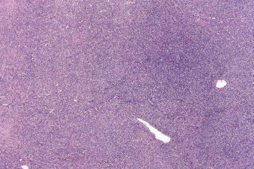
Fig. 35.640 Extraskeletal mesenchymal chondrosarcoma: low-power view showing an intensely cellular tumor.
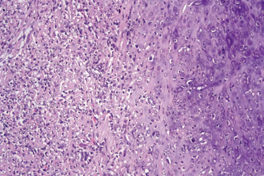
Fig. 35.641 Extraskeletal mesenchymal chondrosarcoma: malignant cartilage is present on the right of the field.