Hibernoma
Hibernoma
Clinical features Hibernoma is a rare, invariably benign tumor resembling normal brown fat, which typically occurs in young adults with a slight female predominance. The tumor most often presents in the interscapular region and thigh followed by the axillae, chest wall and head and neck.1–5 It is a slowly growing, highly vascular lesion that may attain a considerable size and is typified macroscopically by tannish-brown fatty tissue. The large majority of tumors are subcutaneous but about 10% of cases are intramuscular.1 Lesions are benign and there is no tendency for local recurrence.
Pathogenesis and histologic features In cytogenetic studies, hibernomas have shown consistent rearrangement at 11q13.1,6,7 Additionally, MEN1 and/or AIP loss have been described.8,9
Hibernomas are classified histologically into four categories: typical (82% of cases), lipoma-like (7%), myxoid (9%), and spindled cell (2%).1,3,10,11 Typical hibernoma is characterized by an admixture of multivacuolated large adipocytes with central nuclei, large cells with granular eosinophilic cytoplasm and mature univacuolated adipocytes (Fig. 35.33). They are usually encapsulated and lobulated, being subdivided by fine fibrous septa containing numerous small capillaries. Some cases have focal myxoid stroma and very few resemble spindle cell lipoma.1,9 In a subset of lesions large lipoblast-like cells mimicking atypical lipomatous tumors may be conspicuous.12
Tumor cells show variable staining for S100 protein; in the spindle cell variant, the spindled cells are CD34 positive.1 UCP1 positivity has been reported.13
1710 Connective tissue tumors
Differential diagnosis
Distinction from a granular cell tumor is easy, as in the latter the cells are not vacuolated and there are no mature adipocytes. Lesions with lipoblast-like cells have areas that are otherwise typical of hibernoma and tumor cells are negative for MDM2/CDK4.
Adipocytic tumors of intermediate malignancy (locally aggressive)
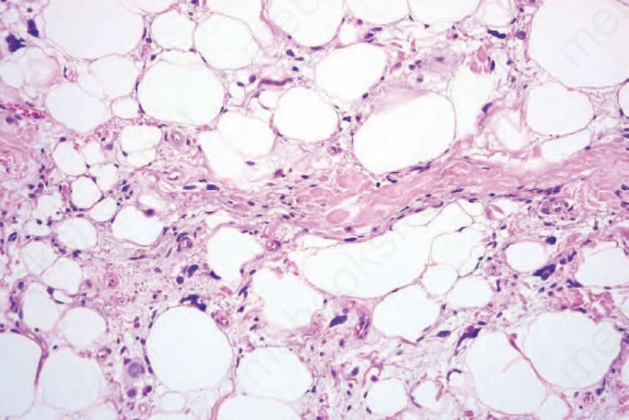
Fig. 35.31 Pleomorphic lipoma: conspicuous floret giant cells are present.
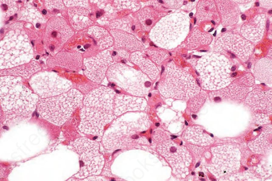
Fig. 35.33 Hibernoma: the admixture of mature adipocytes and large cells with eosinophilic granular cytoplasm is characteristic.
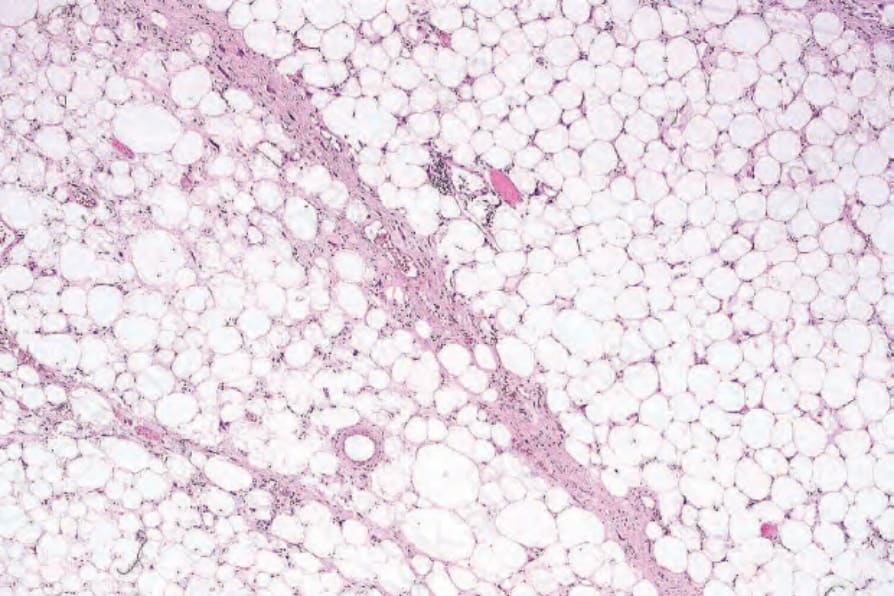
Fig. 35.34 Atypical lipomatous tumor: note the characteristic variation in adipocyte size and the scattered hyperchromatic cells.