Extraskeletal osteosarcoma
Extraskeletal osteosarcoma
Clinical features
Extraskeletal osteosarcoma is a rare lesion that most often arises in older adults and shows an equal sex incidence.1–6 Occurrence in children is unusual.7–10 It typically occurs in the deep soft tissues of the limbs, particularly the legs, but subcutaneous cases are well recognized and exceptional cases are dermal in location.1–6,11–13 Lesion may rarely affect the hand, the breast and the penis.8,14–19 Cases arising on a scar are exceptional.20,21 A single case arising in a lipoma, one in myositis ossificans and a further in a recurrent ossifying fibromyxoid tumor of soft tissue have been reported.22–24
A case of extraskeletal osteosarcoma arising in the mediastinum metastasized to the skin.25
Up to 10% of lesions are associated with previous radiation to the affected site.5,26–30
These tumors tend towards rapid local recurrence and widespread systemic dissemination. The mortality rate is as high as 75%. Tumor size and age of the patient are the most relevant clinical prognostic factors. The role of chemotherapy and/or radiation has not been established.31–33
A better biological behavior, analogous to parosteal osteosarcoma, has been reported for a subset of tumors characterized by 12q amplification.34 In a recent study, potential aggressive molecular subgroups include tumors with CDKN2A loss or biallelic simultaneous losses of RB1 and TP53.35
Pathogenesis and histologic features MDM2 amplification, which correlates with protein immunoexpression, is a not an unusual finding.35–37 Sonic Hedgehog (encoded by SHH gene) and PIK3CA mutations have been documented.35
The tumor is typically ill-defined and characterized by a variable admixture of pleomorphic or spindle-shaped cells associated with the production, at least focally, of an osteoid or chondroid matrix (Figs 35.632–35.634).
1876 Connective tissue tumors
Bizarre multinucleated giant cells are common. The diagnostic sine qua non is the presence of hyperchromatic osteoblasts within the newly formed osteoid matrix. Cases with numerous osteoclastic giant cells were often formerly labeled as so-called giant cell ‘MFH’; such examples not infrequently are subcutaneous. Low-grade variants are exceptional.38 A small cell variant has been described.39–40 Immunohistochemical demonstration of osteocalcin and/or SATB2 may be useful in tumors with poor osteoid formation, whereas ERG may highlight areas with cartilaginous differentiation.41–43
at the periphery of the tumor lobules. These features may be associated with a histiocytic and osteoclastic giant cell reaction. By immunohistochemistry ERG is frequently positive in the cartilaginous component.28
A typical feature, often of diagnostic concern, is the hypercellularity of the lesional cartilage, often with binucleated nuclei and focal nuclear atypia. In the context of a bone tumor, such features would be suggestive of malignancy. Some cases are composed of small, rounded, more primitive chondroblasts, often in a myxoid stroma.29,30
If the presence of a primary lesion in bone has been carefully excluded, the diagnosis of a benign chondroma is assured, despite the worrying features described above. The basis of this assumption is that there is no convincing evidence that a lesion such as an extraskeletal well-differentiated chondrosarcoma exists.
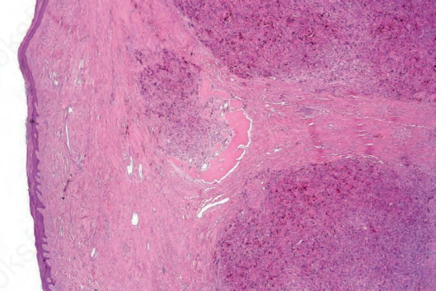
Fig. 35.632 Extraskeletal osteosarcoma: low-power view showing an osteoclast-rich cellular infiltrate with focal osteoid production in the center of the field.
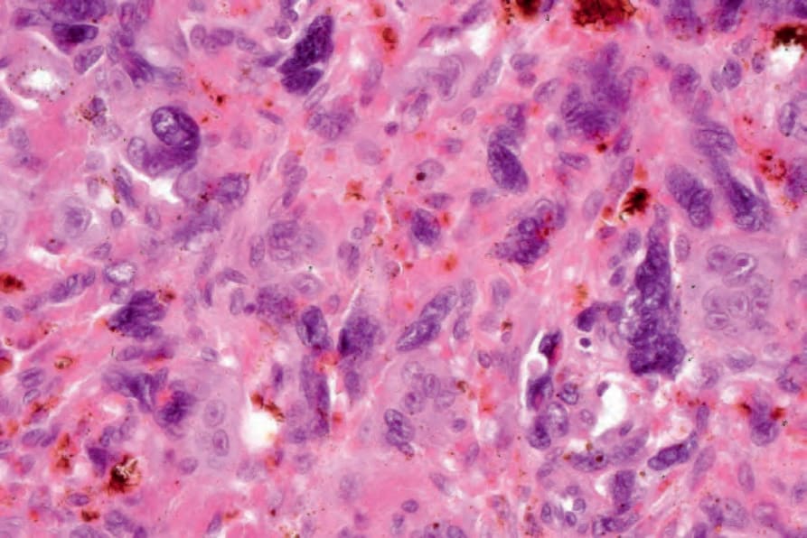
Fig. 35.633 Extraskeletal osteosarcoma: high-power view showing nuclear pleomorphism.
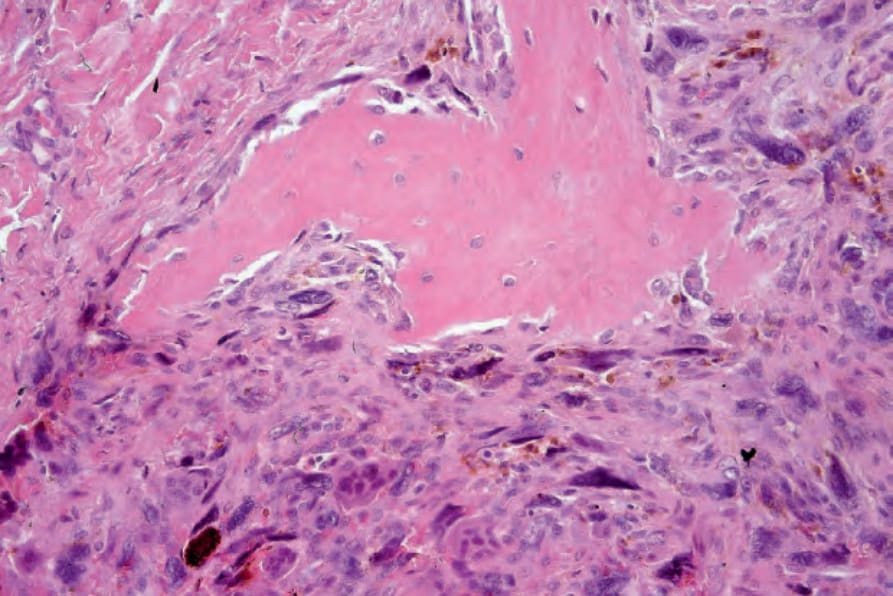
Fig. 35.634 Extraskeletal osteosarcoma: the osteoid is rimmed by malignant osteoblasts.
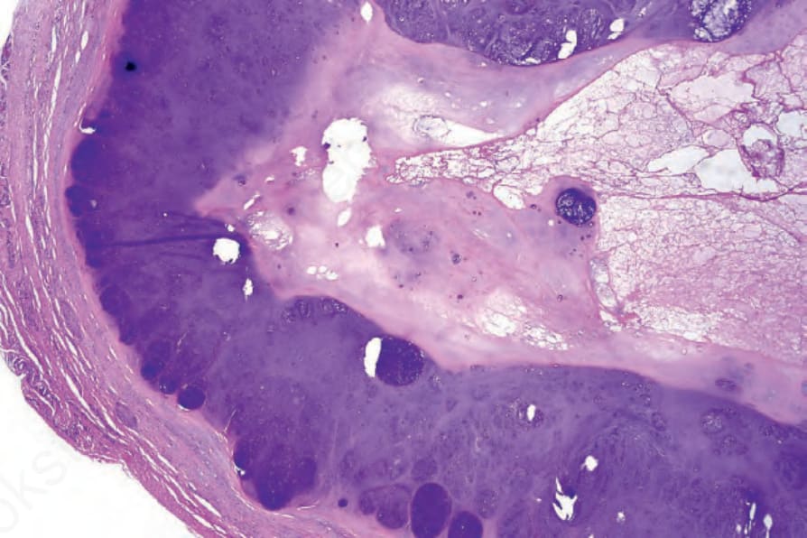
Fig. 35.635 Soft tissue chondroma: the tumor is encapsulated and composed of well-defined lobules of mature cartilage.
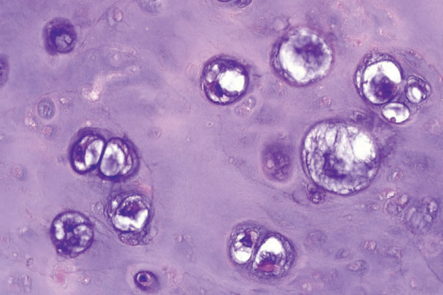
Fig. 35.636 Soft tissue chondroma: the nuclei are typically irregular. In many areas, multiple chondrocytes occupy individual lacunae.