Osteoma cutis
Osteoma cutis
Clinical features Osteoma cutis is a rare benign lesion of the dermis that may be seen at any age in either sex.1–3 Lesions are occasionally multiple and, in some cases, may be inherited or associated with diaphyseal aclasis. Microscopic osteoma cutis is not uncommonly found as a result of dystrophic ossification in association with inflammatory conditions including acne and folliculitis.4,5 The latter is often seen in association with intradermal nevi, as the latter obstruct the hair follicle and lead to inflammation.6 A case of Becker nevus bearing an osteoma cutis has been described.7 Multiple miliary osteoma cutis may occur on the face of middle-aged patients with marked predilection for females.8–13 The lesions are tiny and the etiology is unknown,
A case of pigmented osteoma cutis resulting from long-term tetracycline treatment and a further case after alendronate therapy for osteoporosis has been documented.25,26
A distinctive plaque plate-like variant of osteoma cutis can occur and it is rarely congenital.27–33 It may be multiple and is exceptionally associated with transepidermal elimination.34
Perforating and eruptive and giant cases as well as a case of an extramedullary acute leukemia developing in an osteoma cutis have been reported.18,35–38
Osteoma cutis can be reliably identified in imaging studies sometimes as an incidental finding.39–44
Histologic features Histologically, osteoma cutis is composed of a well-circumscribed nodule of mature lamellar bone, often containing marrow spaces, within the dermis (Fig. 35.631).
1875 Malignant tumors of bone
Differential diagnosis
Osteoma cutis should not be confused with a benign cartilaginous exostosis. The latter condition, also known as osteochondroma, most often presents under the nail as a solitary, often painful, hard tumor nodule. Histologically, it is composed of mature cartilage overlying a layer of lamellar bone. It arises from the underlying phalanx.
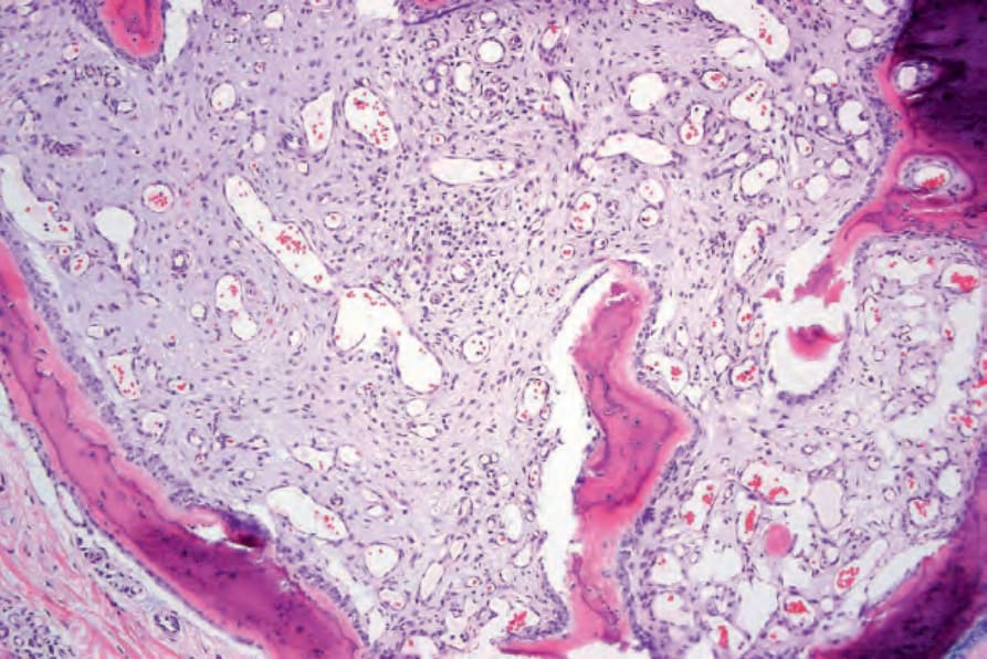
Fig. 35.631 Osteoma cutis: note the osteoid rimmed by osteoblasts and the scarred medullary cavity.
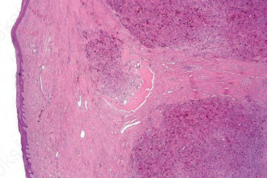
Fig. 35.632 Extraskeletal osteosarcoma: low-power view showing an osteoclast-rich cellular infiltrate with focal osteoid production in the center of the field.