Glomus tumor
Glomus tumor
Glomus tumor arises from the glomus body, which is a specialized arteriovenous anastomosis found most often in the fingers and palms and characterized by the Sucquet-Hoyer canal. They are thought to serve as thermoregulatory receptors. The precise cell of origin is probably a modified smooth muscle cell – the glomus cell – found scattered within the muscle coat of the Sucquet-Hoyer canal.
Clinical features Glomus tumors are relatively common lesions and arise most often in the third and fourth decades, with an equal sex incidence.1–3 They may occur at almost any cutaneous site, but are predominantly seen on the hands,
In a small proportion of cases, the tumors are multiple and may be segmental in distribution.25,26 Multiple lesions are usually seen in children (an otherwise unusual age group) and have an autosomal dominant inheritance (Figs 35.606 and 35.607).27–29 Congenital lesions may also occur and in one there was associated hypertrichosis.30–32 Familial glomangiomas, also known as glomuvenous malformations, are associated with inactivating mutations in GLMN (1p21-22), encoding glomulin which is normally expressed on vascular smooth muscle cells.33–40 Local and systemic expression of basic fibroblast growth factor has been found in occasional patients with multiple glomangiomas, suggesting that this cytokine may play a role in their pathogenesis.41,42
1868 Connective tissue tumors
organoid, mantle of uniformly round glomus cells with pale eosinophilic cytoplasm, clearly defined cell margins and central nuclei (Figs 35.608 and 35.609). Lesional vessels have no demonstrable elastic laminae, and the glomus cells may extend to abut the endothelium or be separated from it by a thin layer of smooth muscle cells. These predominantly solid glomus tumors (the ‘classic’ type) are in fact less common than glomangiomas (see below). Mitotic figures, which are always normal, are only rarely seen and pleomorphism is not a feature. Small nerve fibers may occasionally be demonstrated ramifying through the tumor. Normal blood vessels adjacent to the tumors are usually surrounded by groups of glomus cells. Rare cases may show extensive oncocytic change.54 Prominent epithelioid cell change has been documented (Figs 35.610 and 35.611) and in a single case there was prominent sclerosis.55,56 Calcification is exceptional.57
Multiple or solitary glomus tumors have been described in neurofibromatosis type I.43–45 Tumors tend to occur on the fingers and toes and they are now regarded as part of the spectrum of NF1.46 Glomus tumors in this setting appear to be related to hyperactivation of RAS mitogen-activated protein kinase, resulting from the lack of inhibition by neurofibromin.46
Local recurrence, which is uncommon, only follows inadequate excision and is therefore more frequent in those rare cases (usually deep seated) with infiltrative margins.47 Digital glomus tumors that are skin-colored or those that arise in the nail matrix appear to have a higher risk of local recurrence.48 Rare lesions originate within a blood vessel or a nerve.49–52.The glomus coccygeum, is a prominent glomus body located near the tip of the coccyx that can be found incidentally, and be confused with a neoplasm.53
Histologic features The vast majority of glomus tumors are well circumscribed and composed of small vessels with normal endothelium surrounded by a dense, rather
The tumor cells are positive for SMA, muscle-specific actin and, depending on the antibody, myosin; they are only rarely focally positive for desmin.58 CD34 may also be positive.59 Interestingly, BRAF mutations have been identified in some glomus tumors.60.
In glomangioma, the most common variant of glomus tumor (up to 60% of cases), the vascular component is more prominent and the lumina tend to be somewhat dilated or cavernous (Figs 35.612 and 35.613). Glomus cells may be distributed as an attenuated monolayer or bilayer in the vessel wall.
1869 Tumors of perivascular cells
These cases often exhibit hyalinization of the vessel walls and may show thrombosis with the formation of phleboliths.
• Symplastic glomus tumor is defined as a tumor with high nuclear grade in the absence of any other malignant feature.61–63
• Glomangiomyoma, the rarest subtype (15% of cases), is characterized by a larger and more obvious number of smooth muscle cells, most often distributed adjacent to or around the vascular spaces. These muscle cells merge imperceptibly with the surrounding solid collection of glomus cells (Figs 35.614 and 35.615).
• Glomangiomatosis is defined as a tumor with features of angiomatosis and excess glomus cells.61,64
• Infiltrating glomus tumor is a very rare variant that usually presents in deeper soft tissues.48,65 It is characterized by an infiltrative growth pattern and a high recurrence rate.
• Malignant glomus tumors are rare.48,61,66–70 A single superficial case was associated with pregnancy.71 The histologic diagnosis is difficult and only recently have refined criteria been proposed to define malignant lesions.61 These include:
• deep location and a size of more than 2 cm, or
• atypical mitotic figures, or
• moderate to high nuclear grade diagnosis and five or more mitotic figures per 50 high-power fields (HPF).61
1870 Connective tissue tumors
Glomus tumors of uncertain malignant potential are defined as lesions that lack criteria for the diagnosis of malignant glomus tumor or symplastic glomus tumor but have high mitotic activity and superficial location, or large size only, or deep location only.61 Some 38% of cases fulfilling criteria for malignancy metastasize.61. NOTCH1 and NOTCH3 fusions have been identified in visceral and soft tissue glomus tumors including malignant variants.72
Differential diagnosis The classical clinical history combined with the distinctive histologic features usually prevents diagnostic confusion. Eccrine spiradenoma can be distinguished by the presence in the latter of two populations of cells, positivity for epithelial markers and focal ductal differentiation.
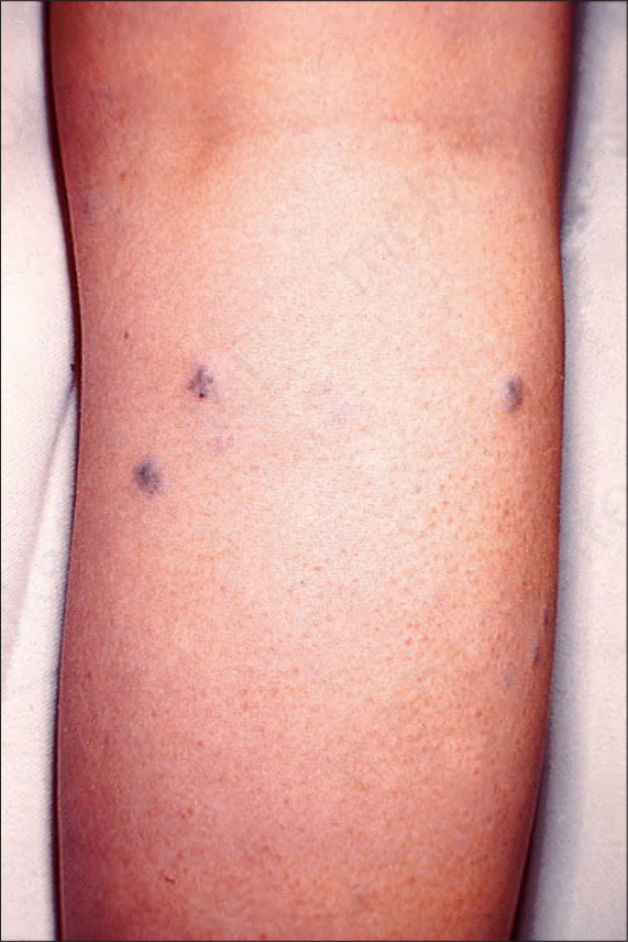
Fig. 35.606 Glomus tumor: multiple, typically small, reddishblue papules are present on the forearm of a young male. By courtesy of the late M. Beare, MD, Royal Victoria Hospital, Belfast, UK.
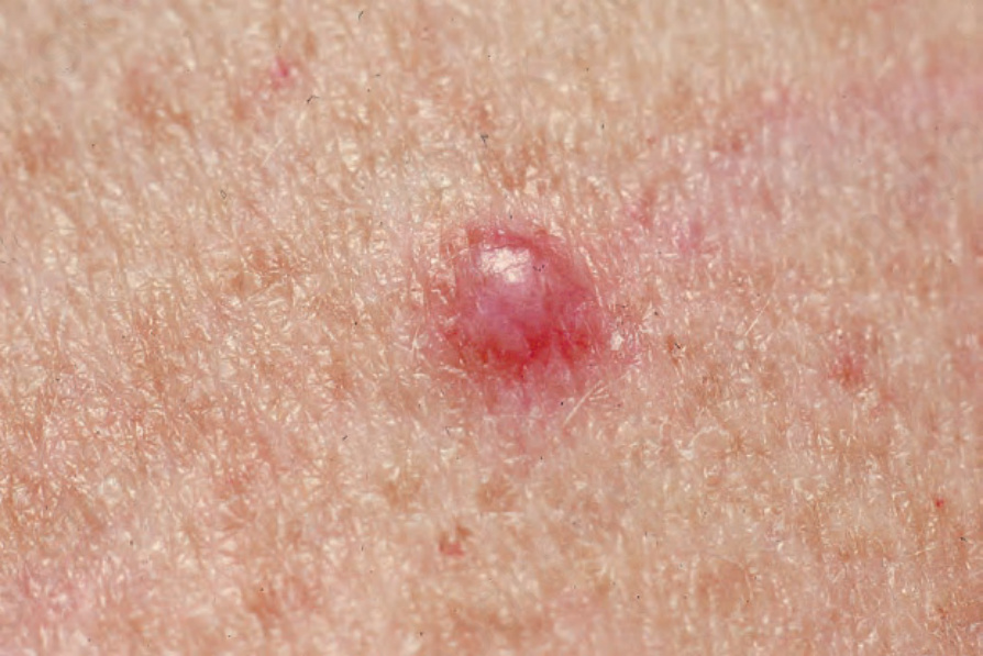
Fig. 35.607 Glomus tumor: close-up view. From the collection of the late N.P. Smith, MD, the Institute of Dermatology, London, UK.
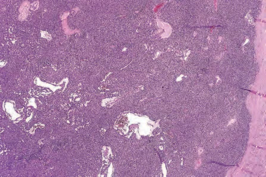
Fig. 35.608 Glomus tumor: the tumor consists of uniform small cells with eosinophilic cytoplasm associated with a conspicuous vasculature.
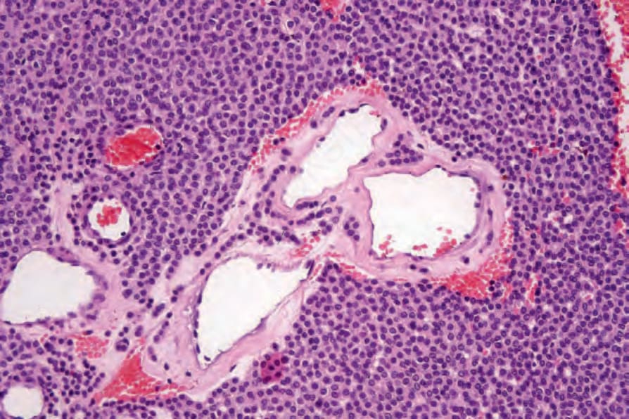
Fig. 35.609 Glomus tumor: the glomus cells have round regular small nuclei. Small numbers are present in this blood vessel wall.
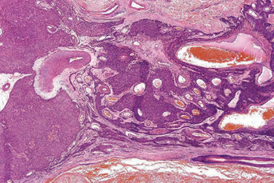
Fig. 35.610 Epithelioid glomus tumor: this field shows the transition between typical small round glomus cells and larger epithelioid variants with abundant pale pink cytoplasm.
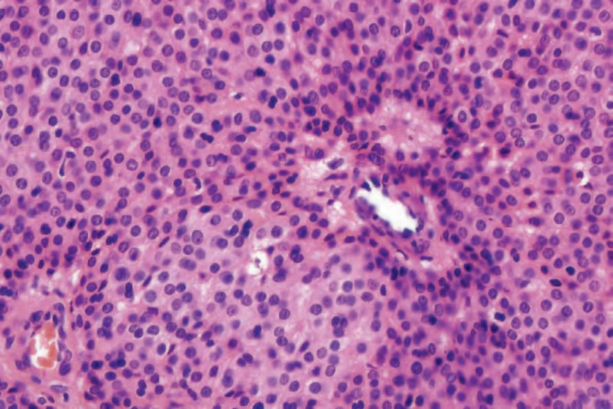
Fig. 35.611 Epithelioid glomus tumor: high-power view.
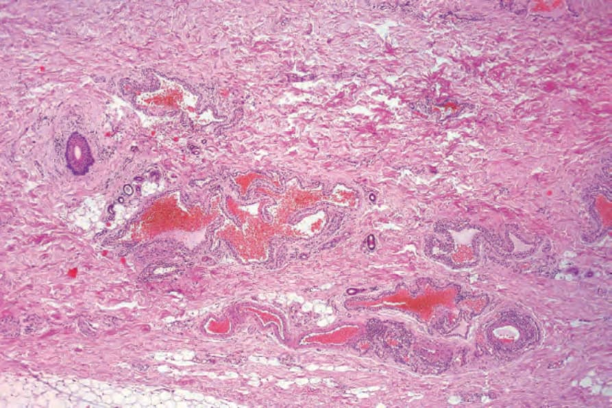
Fig. 35.612 Glomangioma: in this variant, the blood vessels predominate.
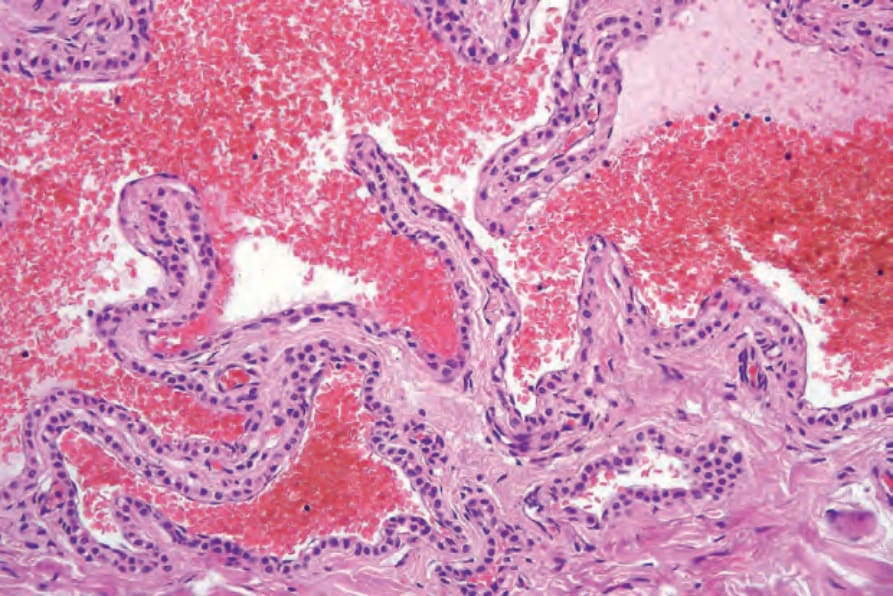
Fig. 35.613 Glomangioma: high-power view.
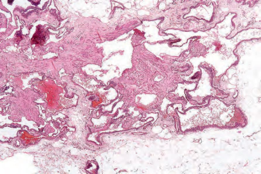
Fig. 35.614 Glomangiomyoma: in this variant, bundles of smooth muscle are present.
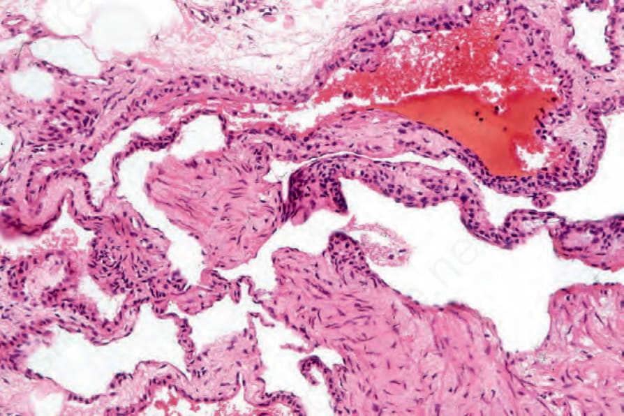
Fig. 35.615 Glomangiomyoma: high-power view.
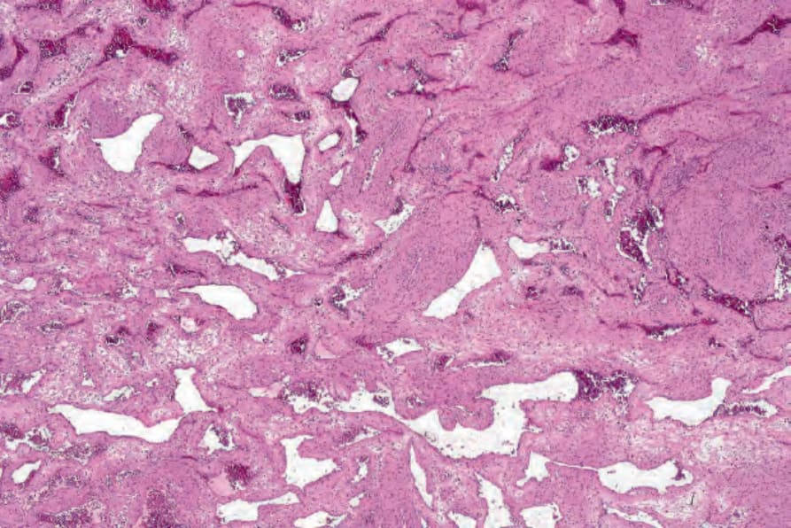
Fig. 35.616 Myopericytoma: low-power view showing dilated vessels and abundant smooth muscle.