Composite hemangioendothelioma
Composite hemangioendothelioma
Clinical features Composite hemangioendothelioma is a low-grade malignant vascular tumor with a tendency for local recurrence but low metastatic potential. It is defined as a neoplasm containing a mixture of histologic patterns including benign, intermediate and/or malignant.1 It is a very rare tumor, presenting mainly
1854 Connective tissue tumors
in adults and only exceptionally in children.1–6 Two congenital cases, one associated with Kasabach-Merritt and one associated with Maffucci syndrome have been reported.3 There is no sex predilection and most tumors occur in the extremities, with a predilection for the hands and feet. A tumor arising in the mediastinum, one on the kidney, one on the spleen, one on the hypopharynx and two in the oral cavity have been documented.3,7–10 In 25% of patients, tumors arise in association with lymphedema and present as long-standing red–blue nodules or plaques. A case presenting with alopecia has been reported.11 The rate of local recurrence is around 50% and this may occur years after excision of the primary tumor. The metastatic rate is around 15%. Rare cases have been reported to metastasize to regional lymph nodes.1,12,13 One of these cases was associated with satellitosis.13 Occasionally, progression to high-grade angiosarcoma can occur over a period of many years.
The prognosis is likely to depend on the component with the highest histologic grade (see below) but this should be confirmed in larger series of cases with adequate follow-up. A handful of cases with expression of neuroendocrine markers and more aggressive behavior have been described.14
Histologic features Composite hemangioendothelioma is a poorly circumscribed dermal and subcutaneous tumor, with an infiltrative growth pattern. The different components vary from lesion to lesion and may include retiform hemangioendothelioma, epithelioid hemangioendothelioma, spindle cell hemangioma, conventional angiosarcoma (low and even high grade), epithelioid angiosarcoma, lymphangioma circumscriptum and areas simulating an arteriovenous malformation (Figs 35.554–35.557).1,3,15 Cases with expression of neuroendocrine markers are characterized by components similar to retiform hemangioendothelioma and epithelioid hemangioendothelioma, and they often have hemangioma-like areas in which channels are lined by hobnail endothelial cells.14 These tumors display synaptophysin, less commonly CD56 and exceptionally, chromogranin.14 Immunohistochemistry displays positive staining for vascular markers including, ERG, CD31 and CD34. D2-40 is positive in a small number of cases.
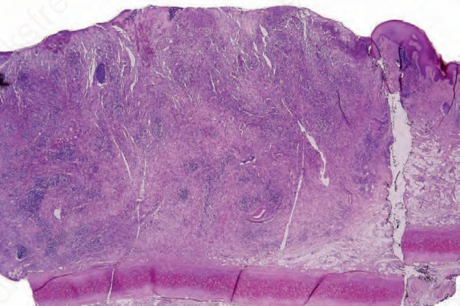
Fig. 35.554 Composite hemangioendothelioma: this lesion is characterized by various vascular patterns.
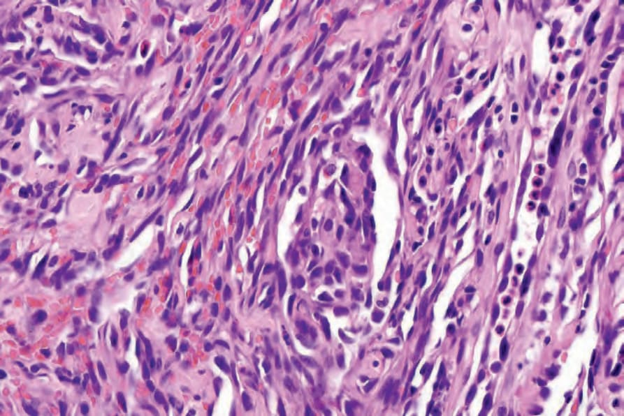
Fig. 35.555 Composite hemangioendothelioma: in this field, the features are reminiscent of spindle cell hemangioma.
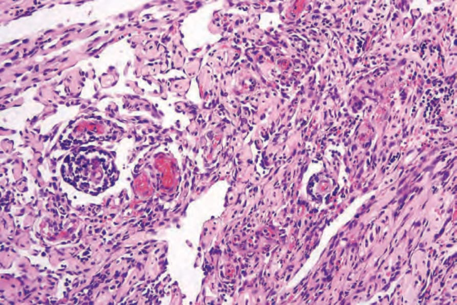
Fig. 35.556 Composite hemangioendothelioma: the appearances resemble papillary intralymphatic angioendothelioma.