Papillary intralymphatic angioendothelioma
Papillary intralymphatic angioendothelioma
Papillary intralymphatic angioendothelioma (PILA) is a very rare tumor, first described by Dabska in 1969 as malignant endolymphatic angioendothelioma (Dabska tumor).1 Since then, only very few additional cases had been reported in the literature, and there has been no consensus regarding its specific histologic features. A recent series has delineated the histologic features of this tumor more accurately and the alternative name of PILA has been proposed.2 Tumors present mainly in infants and children but around 25% of patients are adults.1–3 Males and females are equally affected and tumors have predilection for the limbs. Clinical presentation is that of a slowly growing, solitary, asymptomatic nodule or plaque. Single or multiple tumors have been described in bone.4–7 Exceptional cases have been documented on the spleen, testis and tongue.8–10 In a single case, an angiosarcoma developed within a Dabska tumor.11
Classification as a tumor with low-grade malignant potential is based on reports of local recurrence and rare regional lymph node metastasis in the original series.1 However, follow-up in 8 of the 12 cases recently reported showed no evidence of either local recurrence or distant spread.2 This finding raises the possibility that this tumor is benign but confirmation of these findings is required in larger series with longer follow-up. Until this happens, complete excision of these tumors is advised.
Pathogenesis and histologic features Based on the close interaction between lymphocytes and endothelial cells in Dabska tumor, it has been proposed that the hobnail endothelial cells
1847 Vascular tumors of low-grade or borderline malignancy
Differential diagnosis
The differential diagnosis is the same as that for retiform hemangioendothelioma. Occasionally, lesions that represent otherwise ordinary capillary hemangiomas may display focal papillary tufts which may lead to a confusion with a PILA particularly in small samples.13
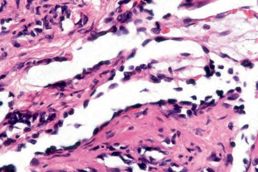
Fig. 35.523 Retiform hemangioendothelioma: intraluminal papillae are commonly present.

Fig. 35.525 Papillary intralymphatic angioendothelioma (Dabska tumor): tumors have dilated, thin-walled vascular spaces mimicking a cavernous lymphangioma. Note the lymphoid aggregates.
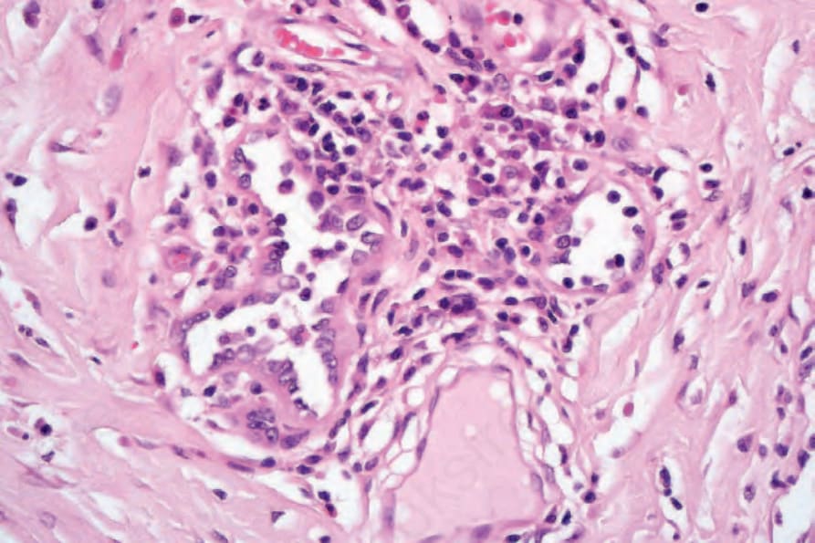
Fig. 35.527 Papillary intralymphatic angioendothelioma (Dabska tumor): hobnail endothelial cells and intraluminal papillae.