Angiomatosis
Angiomatosis
• The second variant consists almost exclusively of capillaries, often with a focal lobular pattern. In both types there is an abundant admixture of mature fat. Glut-1 is negative. Perineural invasion is sometimes a feature. Osseous metaplasia has been reported in one case.6
Clinical features Angiomatosis is a rare condition that presents in children and adolescents.1–3 There is slight predilection for females. A single case involving the left forearm of an adult has been documented.4 It is characterized by a diffuse proliferation of blood vessels affecting a large contiguous area of the body (usually a limb).1,2 Presentation in the head and neck is rarely seen.5 Involvement of the skin, underlying soft tissues and bone is common, and this is associated with hypertrophy of the affected limb. Lesions within parenchymal organs and the central nervous system are sometimes a feature. Due to extensive involvement, surgical treatment is often difficult.
Histologic features Histologically, two patterns have been described:2
• The more common variant consists of a mixture of veins, capillaries and cavernous vascular spaces (Figs 35.515 and 35.516).
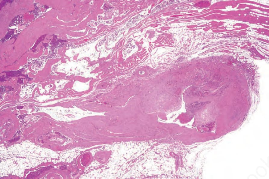
Fig. 35.515 Angiomatosis: this example consists of variably sized congested cavernous vessels.
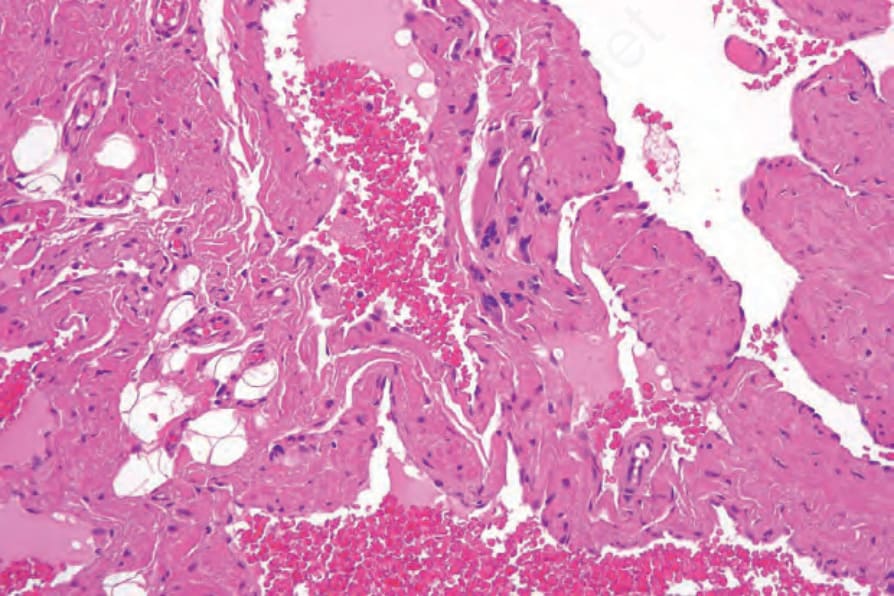
Fig. 35.516 Angiomatosis: high-power view showing dilated vessels with admixed adipocytes.