Myolipoma
Myolipoma
Clinical features Myolipoma, (extrauterine lipoleiomyoma) is a benign adipocytic neoplasm predominantly seen in adult women.1,2 Usually, it presents as an incidental, large (median size 10 cm), deep-seated mass in the retroperitoneum or abdomen and rarely in the pelvis or groin. Rarely, it may be seen as a
smaller palpable subcutaneous lesion in the trunk and extremities.Complete surgical removal is curative.
Pathogenesis and histologic features Fusion of the HMGA2 and C9orf92 genes with t(9;12)(p22;q14) has been reported in one case.3 Myolipoma is well circumscribed with biphasic morphology; variable amounts of mature adipose tissue and well-differentiated smooth muscle fibers arranged in fascicles. Cytologic atypia is not seen. Necrosis is not observed. Unusual features include hypercellular fascicles, degenerative atypia, presence of hemosiderin or eosinophils, metaplastic bone or cartilage, and a round cell component.2 By immunohistochemistry the smooth muscle nature of tumor cells is confirmed by positivity for SMA and desmin. Nuclear positivity of HGMA2 is seen in 2/3rd of cases.
1707 Benign adipocytic tumors and tumorlike lesions
and 35.24). Fibrosis and hemorrhage are often seen. The matrix is composed of chondroitin sulfate.12 Some tumor cells contain glycogen in the cytoplasm. Ossification is exceptional as is the presence of osteoclast-like giant cells.13,14
By immunohistochemistry, mature adipocytes are strongly positive for S100 protein; lipoblasts are only weakly positive for this marker. Chondroid lipomas show strong positivity for Cyclin D1.11 Cytokeratin is very rarely focally positive.2,11,12
Electron microscopy confirms the presence of lipoblasts and mature fat cells. There is no evidence of true cartilaginous differentiation.15
Expression of estrogen and progesterone receptors has been reported.2,4
Differential diagnosis Myolipoma must be differentiated from atypical lipomatous tumor/well-differentiated liposarcoma with smooth muscle differentiation based on the presence of cytologic atypia in the latter.5,6 Moreover, myolipoma is usually negative for MDM2 and CDK4.2 Angiomyolipoma is distinguished by the variable presence of thick-walled vessels and the expression of melanocytic markers, such as HMB45 and Melan-A.6
Differential diagnosis Myxoid liposarcoma usually lacks the presence of a ‘chondroid-like’ matrix and has a characteristic vascular pattern.
In myxoid chondrosarcoma there are no mature adipocytes, although some cells occasionally focally resemble lipoblasts.
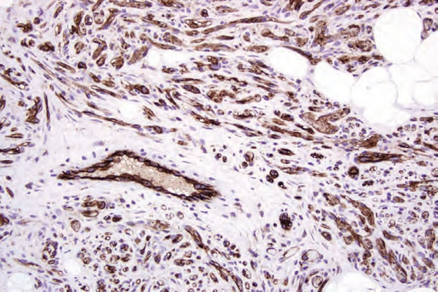
Fig. 35.22 Cellular angiolipoma: the vessels can be outlined with CD31.