Cavernous hemangioma
Cavernous hemangioma
Clinical features The age, sex and anatomical distribution of cavernous hemangioma are much the same as for capillary hemangioma.1–3 However, cavernous hemangioma differs principally in its tendency to be larger and more diffuse, showing little, if any, propensity to involute (Fig. 35.484). These lesions are very likely to be part of the spectrum of lesions described under the rubric noninvoluting congenital hemangioma (RICH, see above). Some of them are also likely to represent vascular malformations. The overlying skin tends towards a rather more bluish-red coloration, reflecting the increased blood content of these lesions.
1836 Connective tissue tumors
Cavernous hemangioma may rarely be associated clinically with multiple enchondromas (Maffucci syndrome), hemangiomas in the alimentary tract (blue rubber bleb nevus syndrome) or with a consumption coagulopathy due to sequestration of platelets within the lesion (Kasabach-Merritt syndrome).4–7
Sinusoidal hemangioma is a relatively uncommon variant of cavernous hemangioma.8 This tumor shares some similarities to a distinctive hemangioma that has been described mainly in the genitourinary tract, soft tissues and other organs as anastomosing hemangioma.9 It presents as a bluish, solitary deep dermal or subcutaneous nodule on the trunk (particularly in the subcutaneous tissue of the breast) or limbs of middle-aged adults, showing a predilection for females. Rare cases in males are associated with gynecomastia. There is no tendency to local recurrence.
Pathogenesis and histologic features It is likely that most cavernous hemangiomas are variants of vascular malformations.
In contrast to capillary hemangiomas, cavernous lesions are composed of a nonlobular, poorly demarcated proliferation of numerous dilated vessels with flattened endothelium (Figs 35.485 and 35.486). Vessel wall thickness is variable. Moderate stromal chronic inflammation is often a feature.
The sinusoidal hemangioma is usually lobular and focally ill defined. It is composed of gaping, markedly dilated, intercommunicating, back-to-back, congested vascular channels with very thin walls, giving rise to a typical sieve-like or sinusoidal pattern (Figs 35.487 and 35.488). Cross-sectioning artifact may produce a pseudopapillary appearance reminiscent of Masson tumor. Focal thrombosis, areas of infarction, hyalinization and even calcification or ossification can be present, especially in long-standing lesions. Endothelial cells are monolayered and flat, but occasionally mild pleomorphism is a feature. Each vessel is surrounded by an attenuated layer of actin-positive pericytes.
Differential diagnosis The diagnosis of sinusoidal hemangioma is usually straightforward, but breast lesions can sometimes be confused with angiosarcoma. The latter, however, is intraparenchymal rather than subcutaneous and shows an infiltrative growth pattern with at least focal endothelial atypia, multilayering and mitoses.
1837 Capillary hemangioma and its variants
layer of SMA positive pericytes. The endothelial cells are positive for CD31, CD34, ERG and WT1 but are negative for podoplanin.3,11,12 GLUT1 is also negative.12,13
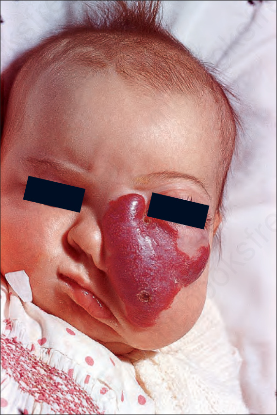
Fig. 35.484 Cavernous hemangioma: this massive lesion is distorting the nose and cheek of this female infant. Cavernous hemangiomas often involve the deeper tissues, with resultant pressure necrosis. By courtesy of M.M. Black, MD, St Thomas’ Hospital, London, UK.
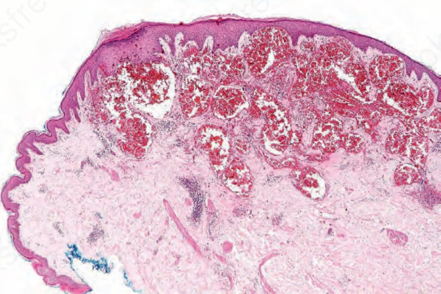
Fig. 35.485 Cavernous hemangioma: the vessels are dilated and rather thin walled.
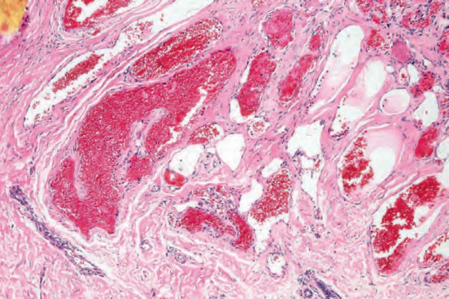
Fig 35.486 Cavernous hemangioma: higher-power view.
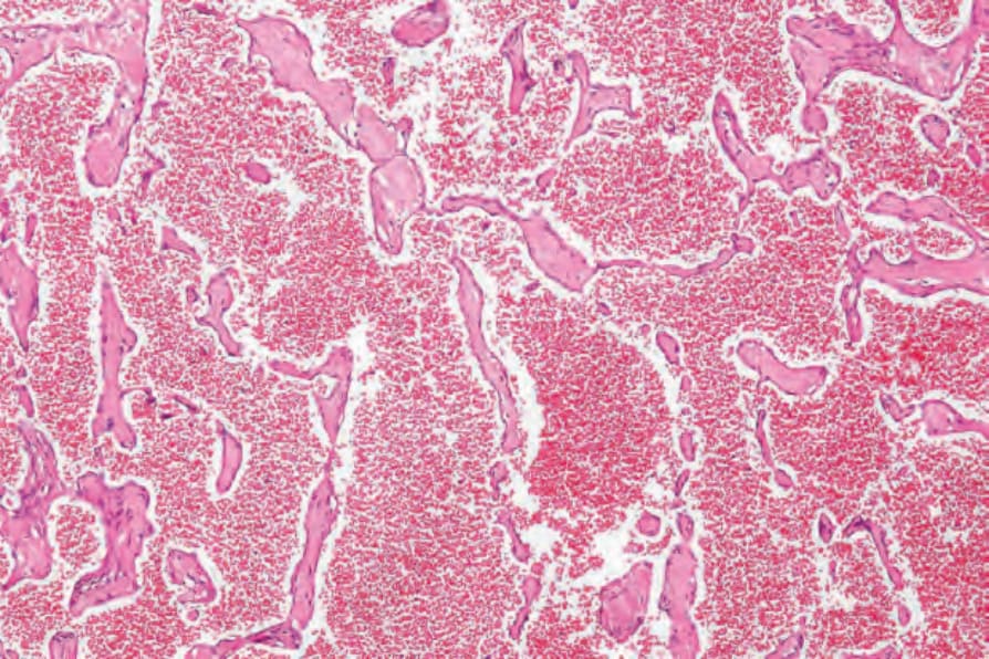
Fig. 35.487 Sinusoidal hemangioma: the back-to-back appearance is characteristic. By courtesy of C.D.M. Fletcher, MD, Brigham and Women’s Hospital and Harvard Medical School, Boston, USA.
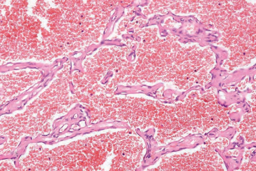
Fig. 35.488 Sinusoidal hemangioma: the presence in some cases of mild nuclear atypia combined with the thin-walled architecture may cause confusion with angiosarcoma. By courtesy of C.D.M. Fletcher, MD, Brigham and Women’s Hospital and Harvard Medical School, Boston, USA.
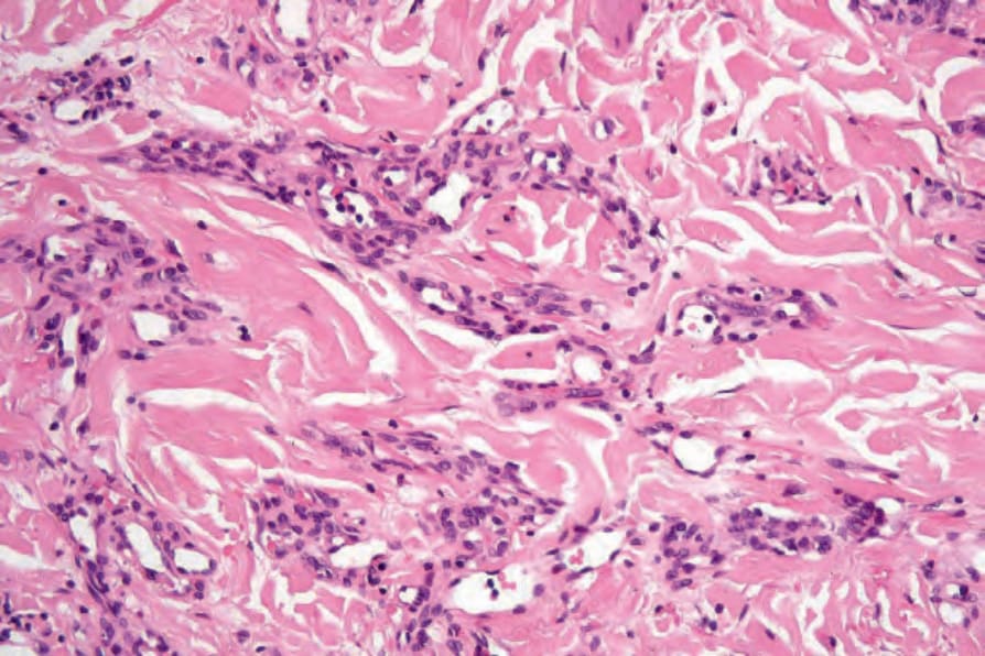
Fig. 35.490 Microvenular hemangioma: the ramifying vessels are lined by a plump endothelial monolayer and an outer layer of more spindled pericytes.