Tufted angioma
Tufted angioma
Clinical features Tufted angioma (angioblastoma of Nakagawa) is a distinctive variant of capillary hemangioma which was described in the Japanese literature in 1949 as angioblastoma.1–4 It presents most commonly in infants or children, showing an equal sex incidence and a predilection for the neck, upper trunk and limbs (Fig. 35.480).1–8 Lesions tend to be acquired mainly during the
1835 Capillary hemangioma and its variants
first year of life, but congenital tumors occur in about 25% of patients.8,9 A congenital case with disseminated lesions has been described.10 Isolated cases occur in adults.11,12 Familial tumors are exceptional.13 Unusual locations of the tumor include the oral cavity, the perianal area and the palm.14–16 A case in an intracranial location, one in the maxilla and one in the nasal cavity have been described.17–19 The lesion grows slowly over a period of years as an erythematous macule or plaque, or as a cluster of papules attaining a size of up to 10 cm or more. An annular configuration can also be seen and multifocal lesions are rare.20–22 In some cases hyperhidrosis and hypertrichosis are seen.8 Kasabach-Merritt syndrome is an important complication in a very small number of cases and more rarely low-grade coagulopathy is seen.8,23–25 Exceptional cases associated with vascular malformations, one occurring at the site of herpes zoster and one at a site of BCG vaccination have been documented.26–29 A case of an angiomatous lesion developing in association with Ramucrimab therapy and histology mimicking tufted angioma was observed.30 Surgical excision is difficult due to the size and extension of the tumor beyond the evident clinical margins. Spontaneous regression does occur, it is usually not complete and seems more common than previously thought.31–34 Regression may even be seen in congenital tumors.35
Histologic features At low power, the distinctive feature is the presence in the dermis and superficial subcutis of scattered, rounded, oval or elongated lobules of closely packed capillaries in a typical ‘cannonball’ distribution (Fig. 35.481). Each lobule is composed of poorly canalized capillaries lined by bland endothelial cells and surrounded by pericytes, and closely resembling the early stages of a strawberry nevus. A distinctive feature is the presence in the periphery of the lobules of dilated crescent-shaped or semilunar lymphatic channels (Fig. 35.482). Focal crystalline inclusions in the endothelial cell cytoplasm can be seen in some cases (Fig. 35.483).36 In the dermis between the tufts of capillaries, variable number of dilated lymphatics are seen.37 Unusual histologic features include an intravenous location and proliferation of sweat glands.38–40 In a case with regression an increase in CD8+ lymphocytes within tumor lobules suggested a cytotoxic mediated immune response as a possible mechanism.41 Areas of tufted hemangioma may occur in kaposiform hemangioendothelioma and it is likely that both tumors are part of the same spectrum.42–44 By immunohistochemistry, the endothelial cells lining the capillaries are positive for ERG and CD31. The crescent-like vessels and the lymphatics in the surrounding dermis are positive for D2-40.45 WT1 is positive.
Differential diagnosis Strawberry nevus has a more diffuse, confluent and extensive involvement of the dermis and subcutis, and lacks the dilated crescent-shaped lymphatic channels at the periphery of the lobules. Distinction from nodular Kaposi sarcoma is easy because the latter lacks the ‘cannonball’ pattern and is composed of a uniform population of spindle-shaped cells and pseudovascular clefts. Furthermore, cutaneous involvement by Kaposi sarcoma in immunocompetent children is exceedingly rare.
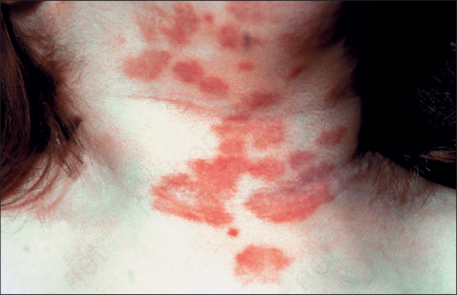
Fig. 35.480 Tufted angioma: lesions commonly present on the neck and upper trunk. Note the presence of extensive macules and plaque-like lesions. By courtesy of the Institute of Dermatology, London, UK.
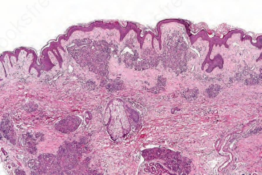
Fig. 35.481 Tufted angioma: this is a typical low-power appearance of sharply circumscribed vascular nodules in the reticular dermis.
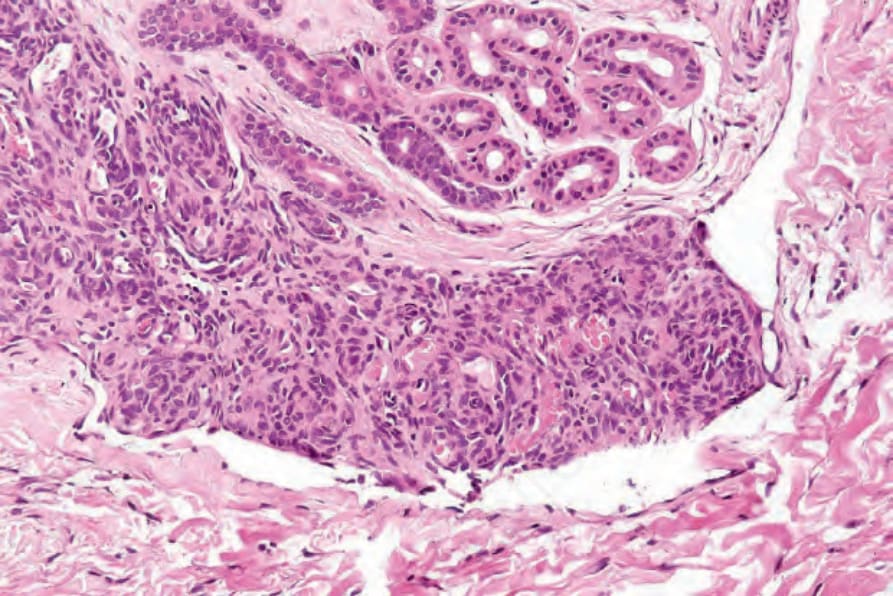
Fig. 35.482 Tufted angioma: the nodules are composed of tightly knit capillaries. Note the conspicuous lymphatic vessel.
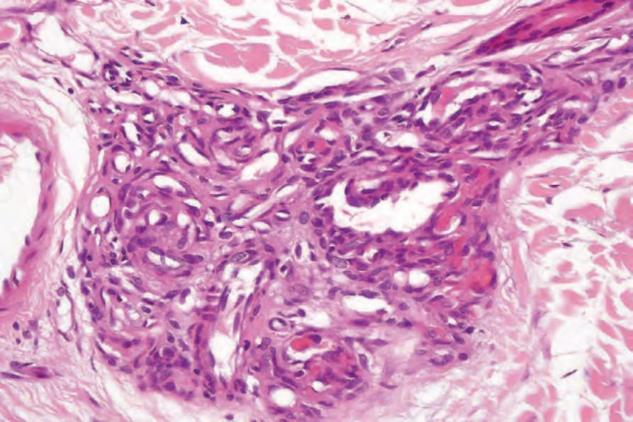
Fig. 35.483 Tufted angioma: note the eosinophilic inclusions.