Angiolipoma
Angiolipoma
Clinical features Angiolipomas are benign lesions which, in contrast to simple lipomas, are seen most often in young adults and have a predilection for the subcutis of the upper limbs, particularly the forearm and less commonly the trunk.1–3 Oral,
1706 Connective tissue tumors
intra-articular, extradural, breast, and bronchial lesions are exceptional.4–8 Familial cases are rarely seen.9 Parenchymal, CNS and infiltrating intramuscular lesions are best regarded as variants of hemangiomas.10
The lesions are typically tender or painful, less than 2 cm in diameter, and may impart a reddish or bluish discoloration to the overlying skin. They are more often multiple than solitary and treatment can be problematic. Multiple lesions have exceptionally been documented in association with diabetes mellitus and as a complication of antiretroviral therapy (particularly with indinavir and saquinavir) in acquired immunodeficiency syndrome (AIDS).11–14 A case of intravascular lymphomatosis as well as that of a B-cell lymphoma presenting in an angiolipoma have been documented.15,16 Metastatic melanoma within an angiolipoma has also been described.17 Giant angiolipomas are very rare.18
Pathogenesis and histologic features Most cytogenetic studies in angiolipoma have consistently shown a normal karyotype.19 In only a few tumors cytogenetic abnormalities have been demonstrated including a one with 46,Y,t(X;2)(p22;p12), one with complete loss of a chromosome 13 and two with structural aberrations of chromosome 13 involving the q12~q14 bands.20,21 Interestingly, more recently, low-level mutations of protein kinase D2 was demonstrated in 80% of angiolipomas.22 Aberrant expression of HMGA2 has been documented.23
The tumor, almost always encapsulated, is composed of mature adipocytes and varying proportions of irregular, anastomosing small blood vessels without endothelial atypia (Fig. 35.19). Luminal microthrombi are invariably present (Fig. 35.20). Although the blood vessels are usually seen in the periphery of the tumor, they may constitute most of the lesion. Such examples are known as cellular angiolipomas (Figs 35.21 and 35.22).24–26
Differential diagnosis Cellular angiolipoma can be confused with a vascular tumor, especially immature capillary hemangioma and kaposiform hemangioendothelioma.17 The presence of mature adipocytes and capillaries with microthrombi allows distinction from immature capillary hemangioma. Kaposiform hemangioendothelioma may have capillaries with microthrombi in the periphery of tumor lobules, but mature adipocytes are absent.
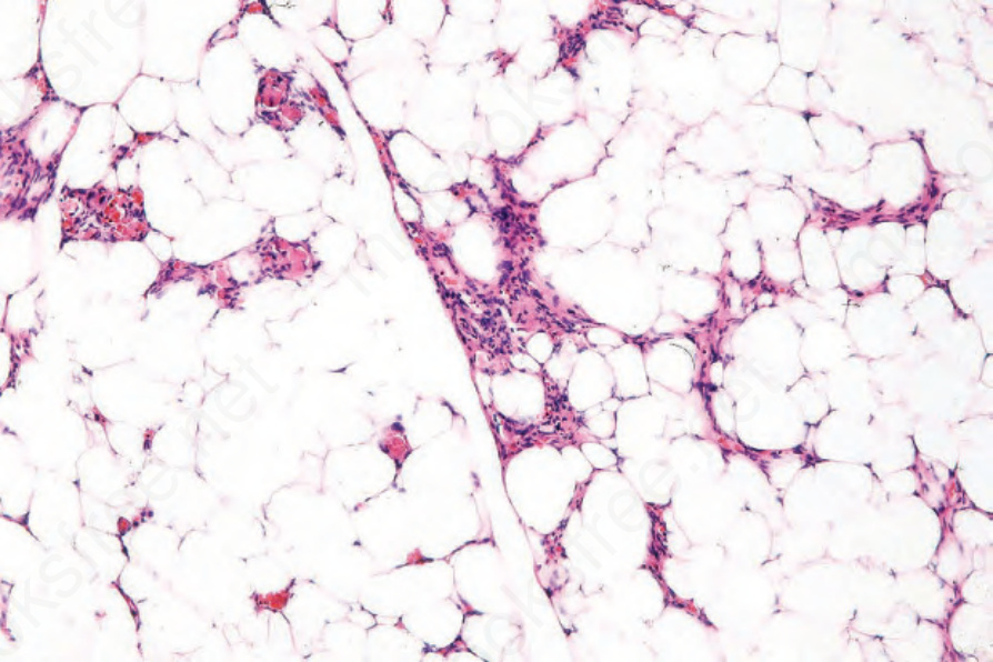
Fig. 35.19 Angiolipoma: admixed with the adipocytes are aggregates of small vessels.
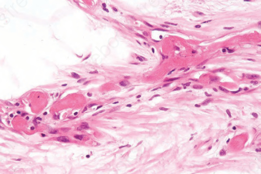
Fig. 35.20 Angiolipoma: vascular thromboses are an invariable feature. They are often found at the periphery of the tumor.
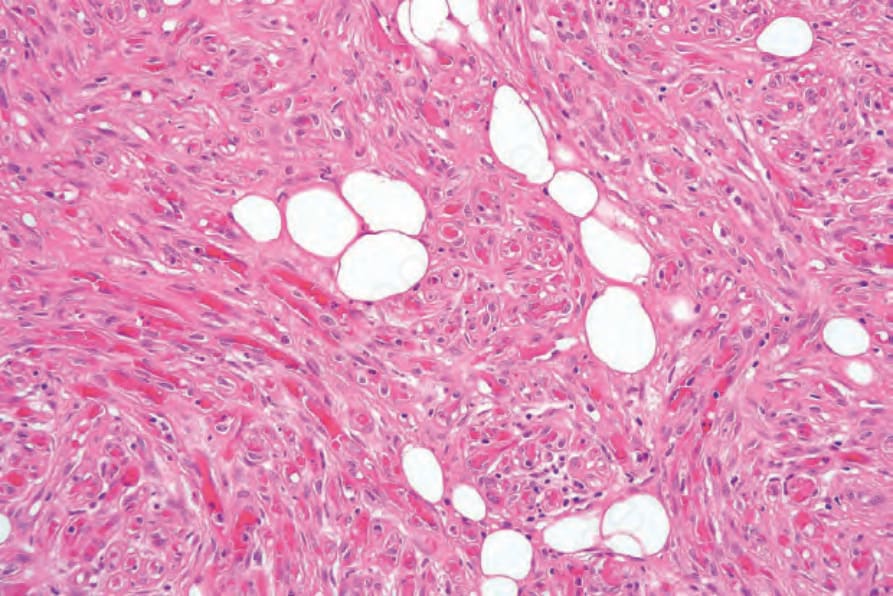
Fig. 35.21 Cellular angiolipoma: in this variant, bland spindled cells (possibly pericytes) predominate. The presence of adipocytes and capillary microthrombi confirms the diagnosis.