Angioma serpiginosum
Angioma serpiginosum
Clinical features Angioma serpiginosum is rare, usually arises in childhood, and occurs particularly on the extremities.1–3 It is characterized by multiple tiny punctate red or purple lesions about the size of a pinhead, typically arranged in a gyrate or serpiginous pattern (Figs 35.455 and 35.456).1–3 New papules tend to form gradually, thereby expanding the lesion. Sometimes the condition may simulate purpura.4 A linear arrangement may rarely be seen.5,6 Ocular and nervous system involvement has occasionally been documented.7,8 Although most cases are localized, extensive involvement rarely occurs.9 Occasional cases are familial.10 In cases with a systematized segmental distribution, genetic mosaicism has been suggested.11
In two-thirds of cases, associated abnormalities – including nevus flammeus, macrocephaly, syndactyly, hydrocephalus, body asymmetry, anal atresia, hearing loss, cardiovascular abnormalities, strabismus, hypothyroidism, nevus anemicus, café-au-lait spots, lipoma and hypospadias – may be seen.1,2,4,5 A subset of patients with cutis marmorata telangiectatica congenita were thought to be associated with macrocephaly, however, nowadays is well known that the capillary malformations in these patients represent a reticulated type of capillary malformations.6 In the Adams-Oliver syndrome, aplasia cutis congenita and transverse limb defects are associated with other malformations in addition to cutis marmorata telangiectatica congenita.7,8
Histologic features Biopsy shows dilatation of capillaries and venules in the superficial dermis. Occasionally, a true vascular proliferation may be seen.9
Pathogenesis and histologic features It has been suggested that the disease is caused by deletions or mutations on chromosome Xp11 encoding for the PORCN gene.12,13 However, this view has been challenged with the suggestion that the patients described had focal dermal hypoplasia.14
1827 Benign tumors including reactive vascular proliferations, malformations and ectasias
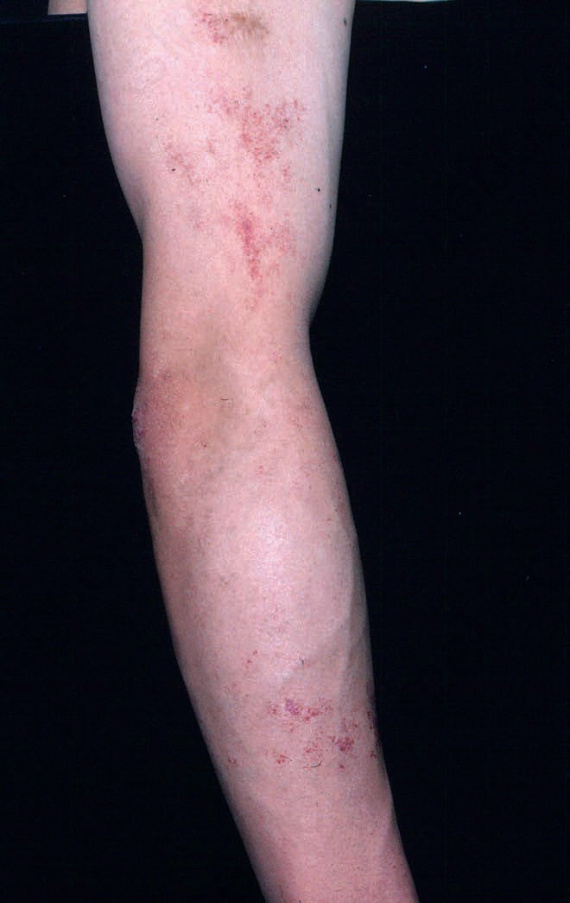
Fig. 35.455 Angioma serpiginosum: the distribution of these tiny red macules is characteristic. From the collection of the late N.P. Smith, MD, The Institute of Dermatology, London, UK.
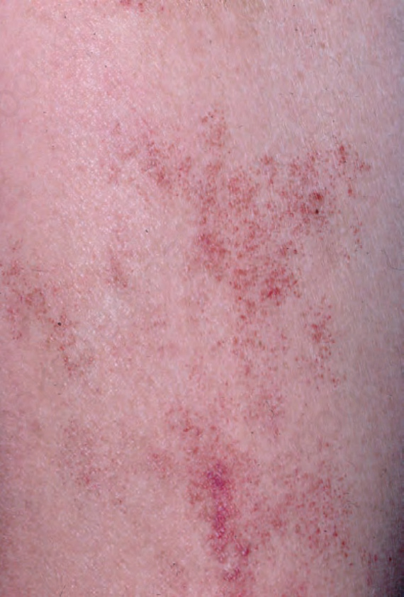
Fig. 35.456 Angioma serpiginosum: close-up view. From the collection of the late N.P. Smith, MD, The Institute of Dermatology, London, UK.
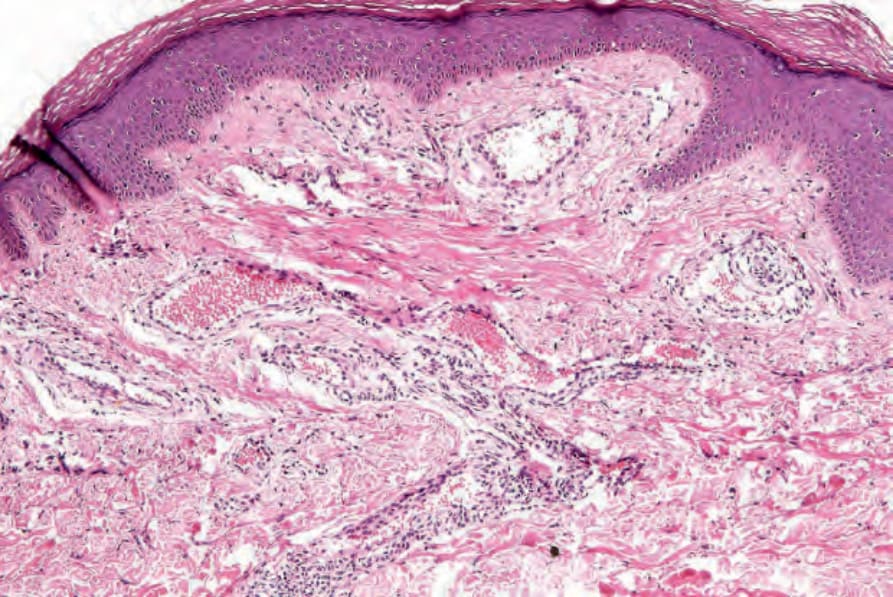
Fig. 35.457 Angioma serpiginosum: histologically, it is composed of a localized cluster of thick-walled and dilated capillaries, usually in the superficial dermis.
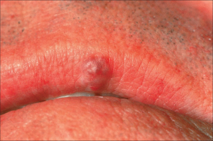
Fig. 35.458 Venous lake: there is a typical blister-like vascular lesion. By courtesy of the Institute of Dermatology, London, UK.
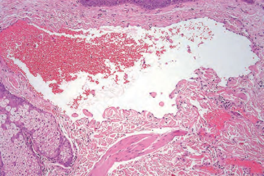
Fig. 35.459 Venous lake: there is striking venous ectasia.