Nevus flammeus
Nevus flammeus
Clinical features The old term nevus flammeus encompasses both the salmon patch and the port-wine stain.1–3. However, at present, the term nevus flammeus is only used to refer to port-wine stains.
Salmon patch Salmon patch (nevus simplex, “angel kiss,” “stork bite) is a congenital lesion that usually presents on the head and neck (forehead and nape of neck) as a reddish-purple macule and tends towards spontaneous involution. It can be seen in up to one-third of neonates, representing the most
1825 Benign tumors including reactive vascular proliferations, malformations and ectasias
common form of vascular malformation.4 In a study of cutaneous findings in hospitalized neonates, a salmon patch was found in 91.2% of patients.5 In a further study in 500 newborns, a salmon patch was found in 28% of full-term infants and in 25.8% of pre-term infants.6 A salmon patch on the nape of the neck seems to be more commonly associated with a mother greater than 35 years of age.7
Port-wine stain Port-wine stain is a lateralized dark lesion that tends towards continued growth and only very rarely involutes (Fig. 35.449).1–3,8 A similar lesion characterized by light-red or pale-pink color has been described as nevus roseus.9 Rare acquired cases of port-wine stain may also be seen, often in association with trauma.10,11
Pathogenesis and histologic features
A somatic activating mutation in the GNAQ gene, located on chromosome 9, has been identified in both syndromic and non-syndromic port wine stains.29 It has been proposed that the endothelial cells of port-wine stains
The familial combination of port-wine stain and arteriovenous malformation appears to be related to mutations in RASA1 (5q13.3) encoding a regulator of the Ras protein.12–15
Port-wine stain may be associated with ipsilateral cerebral or meningeal vascular lesions and vascular eye abnormalities (Sturge-Weber syndrome) or with hypertrophy of a limb and partial venous agenesis with varicosities (Klippel-Trenaunay syndrome)16,17 (Figs 35.450–35.452). In Cobb syndrome, there is a port-wine stain overlying an underlying spinal cord vascular malformation in the midline of the back.18,19 The cutaneous vascular lesion may also represent a verrucous hemangioma.19
Other vascular lesions, particularly lobular capillary hemangioma (pyogenic granuloma), vascular malformations, a combination of the latter two and even tufted angioma, may occur within a port-wine stain.20–25 Lobular capillary hemangioma-like (pyogenic granuloma-like) lesions can occur after laser treatment.26 Basal cell carcinoma may occasionally develop within a port-wine stain.27,28.
1826 Connective tissue tumors
represent differentiation impaired late-stage endothelial progenitor cells that form immature venule-like structures. As a result of disruption in the interaction between endothelial cells in these vessels (co-expressing Eph receptor B1 and ephrin B2), there is progressive dilatation resulting in the abnormality observed.30
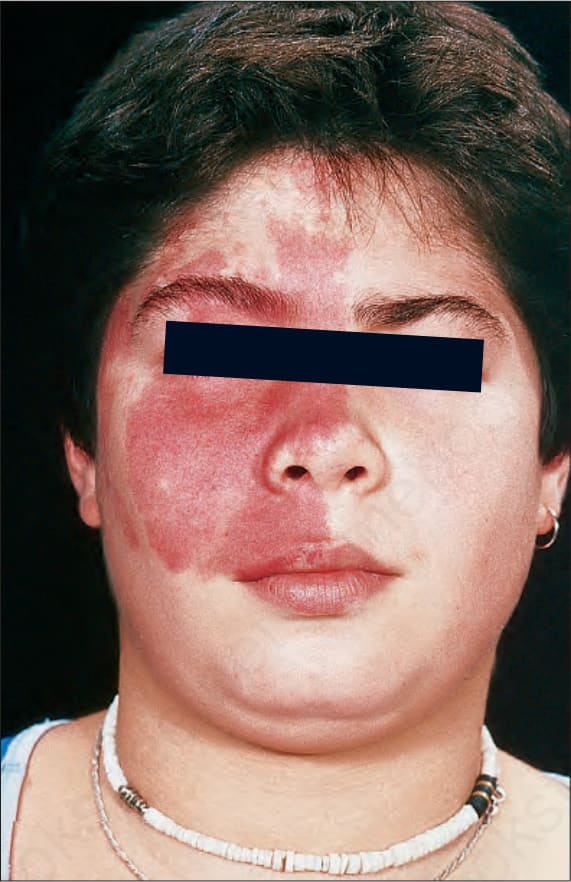
Fig. 35.450 Sturge-Weber syndrome: this 12-year-old girl presented with fits, mental retardation, and a port-wine vascular nevus affecting much of the right side of her face. CT scan showed meningeal angiomatosis. By courtesy of D. Atherton, MD, Institute of Dermatology and Children’s Hospital at Great Ormond Street, London, UK.
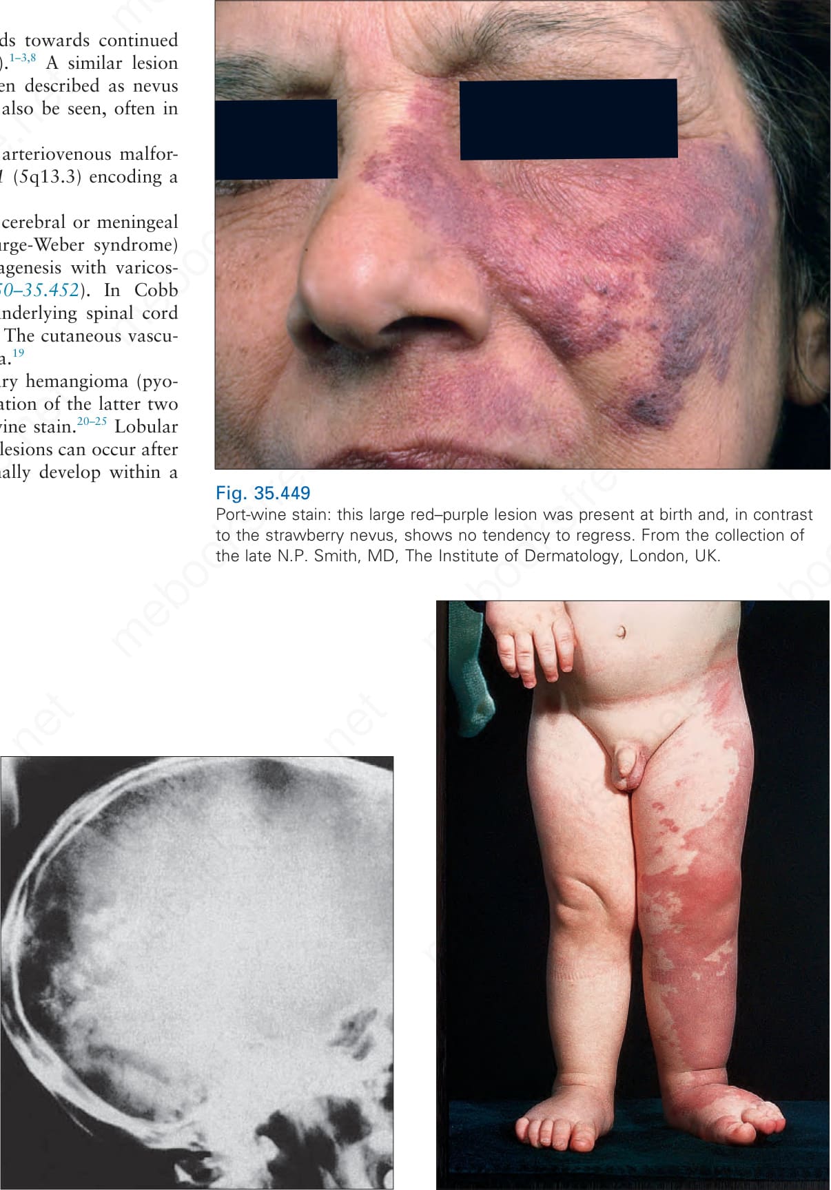
Fig. 35.451 Sturge-Weber syndrome: the intracranial moiety is often calcified, as in this radiograph. By courtesy of I. Moseley, MD, National Hospital for Nervous Diseases, London, UK.
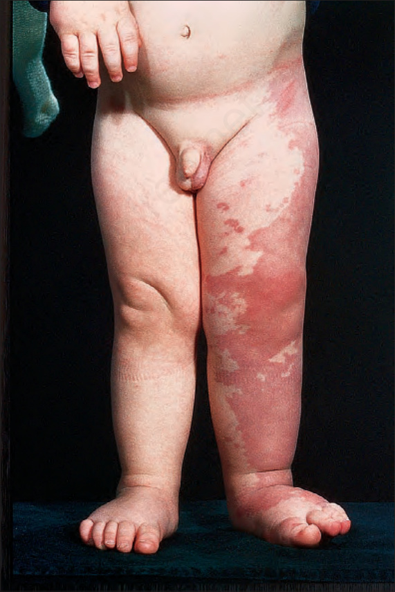
Fig. 35.452 Klippel-Trenaunay syndrome: this 2-year-old boy has a port-wine vascular nevus, affecting much of the skin of the left leg, associated with increased soft tissue growth in the leg and a slight increase in its length. By courtesy of D. Atherton, MD, Institute of Dermatology and Children’s Hospital at Great Ormond Street, London, UK.
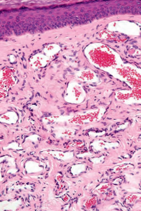
Fig. 35.453 Port-wine stain: the malformation is characterized by numerous dilated blood-filled capillaries.
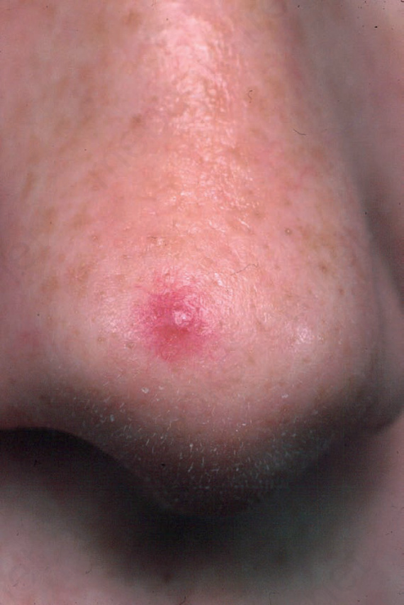
Fig. 35.454 Spider nevus: note the central macule with radiating vessels. From the collection of the late N.P. Smith, MD, The Institute of Dermatology, London, UK.
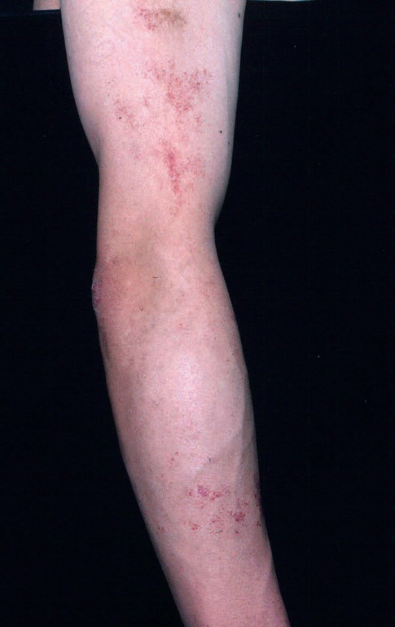
Fig. 35.455 Angioma serpiginosum: the distribution of these tiny red macules is characteristic. From the collection of the late N.P. Smith, MD, The Institute of Dermatology, London, UK.
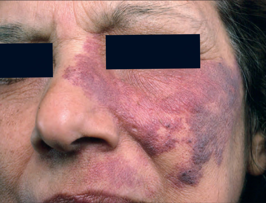
Fig. 35-449 (caption embedded in image / 圖說烘焙於圖內)