Rhabdomyoma (extracardiac)
Rhabdomyoma (extracardiac)
Clinical features Extracardiac rhabdomyomas are very uncommon, usually deep-seated lesions and therefore rarely present to the dermatologist.1–4
Pathogenesis and histologic features Genital type A proliferation of spindle or strap-shaped cells with abundant eosinophilic cytoplasm containing cross striations embedded in a fibrous stroma is typically seen. Pleomorphism or mitotic activity are not seen. A sclerosing variant characterized by a dense collagenous stroma has been identified mainly in males in a paratesticular location, and rarely in women.8,10
Adult type A case with a t(15;17)(q24;p13) has been reported.5
The adult type is composed almost entirely of large, round, polygonal or strap-shaped cells with plentiful eosinophilic cytoplasm (rhabdomyoblasts)2
1819 Malignant striated muscle tumors
(Fig. 35.428). Cross-striations are readily identifiable and rod-like inclusions are commonly present (Fig. 35.429). Oncocytic change is exceptional.39
Differential diagnosis Distinction of any of these lesions from rhabdomyosarcoma is made possible by their lack of mitotic activity, nuclear pleomorphism or infiltrative growth pattern.
Immunohistochemistry shows positivity for muscle-specific actin, myoglobin and desmin.2 Very focal positivity for S100 protein and SMA may also be seen.2
Fetal type Aberrations at the PTCH1 locus suggesting a role for the activated Hedgehog signaling in the pathogenesis of fetal rhabdomyoma have been documented.40
The fetal type is composed almost entirely of immature round to spindle-shaped rhabdomyoblasts in a myxoid stroma, showing progressive maturation towards more eosinophilic cells (which often show cross-striations) peripherally (Figs 35.430 and 35.431).
Immunohistochemistry shows positivity of tumor cells for muscle-specific actin, myoglobin and desmin.3 Focal positivity for SMA, GFAP and S100 protein may be evident.3
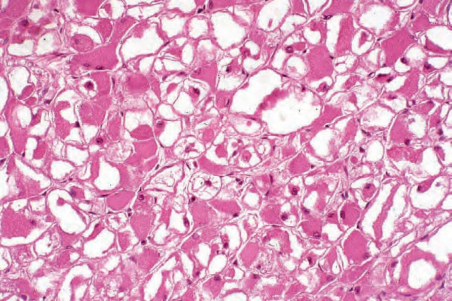
Fig. 35.428 Rhabdomyoma: the adult-type lesion is composed of large polygonal cells with copious eosinophilic cytoplasm and peripherally located nuclei without pleomorphism.
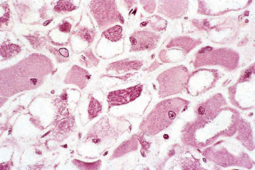
Fig. 35.429 Rhabdomyoma: in the center of the field are pathognomonic ‘jack straw’ intracytoplasmic inclusions.
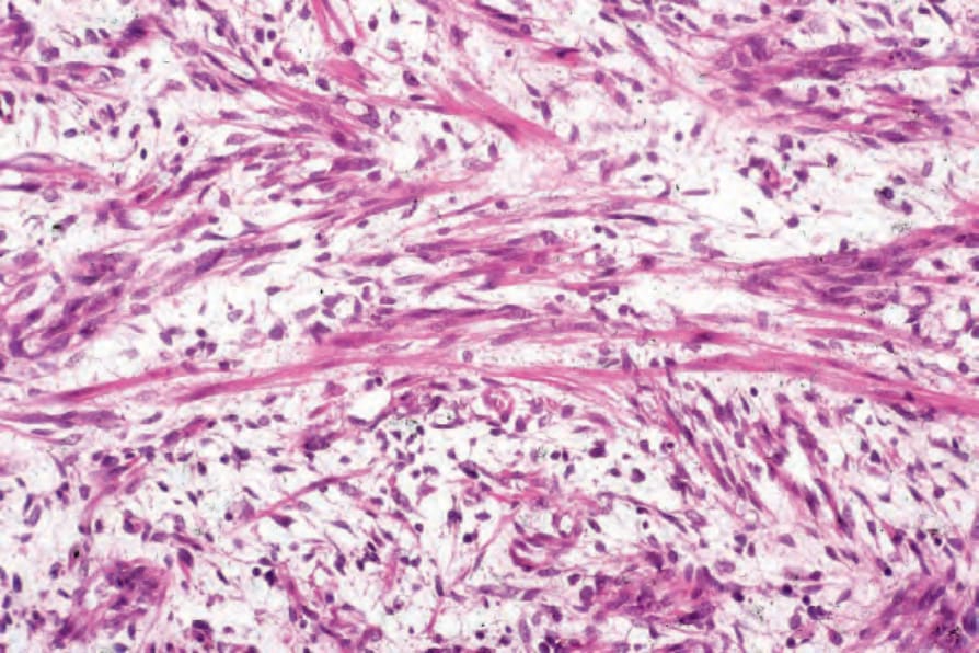
Fig. 35.430 Fetal rhabdomyoma: this example of the fetal myxoid type is composed of obvious elongated strap cells set in a loose myxoid stroma.
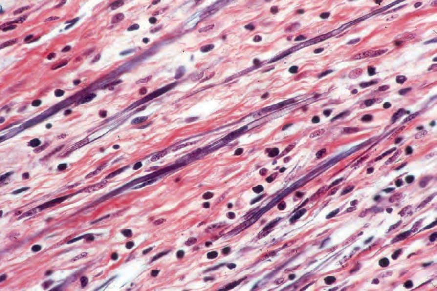
Fig. 35.431 Fetal rhabdomyoma: typical cross-striations are evident (phosphotungstic acid–hematoxylin).