CIX-rearranged sarcoma
CIX-rearranged sarcoma
Clinical features CIX-DUX4 sarcoma is the most common variant of the EWS negative round cell tumors.3–17 It usually occurs in soft tissue, including superficial locations, particularly in trunk and extremities of young adults with no sex predilection.8,15 Children may be affected.6 The clinical course is aggressive and metastases, including lymph nodes are frequent.
Pathogenesis and histologic features DUX-4 transcribes a DNA binding site that upregulates the ETS family genes.9 In addition to DUX4, fusions with FOXO4 and NUTM2A have been documented.3,13
Pathogenesis and histologic features Cytogenetic studies in soft tissue melanoma have shown a specific t(12;22) (q1314;q1213).30–33 As a result of this translocation, there is fusion of the Ewing sarcoma (EWSR1) oncogene and the activating transcription factor 1 (ATF1). Expression of the melanocyte-inducing factor – microphthalmia transcription factor (MITF) – is directly induced by the EWSR1/ ATF1 fusion protein.34–36 CREB1 t(2;22)(q33;q12) can substitute for ATF1 in a small subset of cases.37,38 Fusions of EWSR1 and CREB1 may be more common in the gastrointestinal form of clear cell sarcoma and might be less efficient in promoting melanocytic differentiation.39 Identical gene fusions are seen in angiomatoid fibrous histiocytoma, but MITF transcription is not induced.40,41 Gene expression profiles confirm strong melanocytic differentiation in clear cell sarcoma.42,43 Other neoplasms with the same gene fusions include gastrointestinal (neuroectodermal) clear cell sarcoma (which some consider a distinctive form), angiomatoid fibrous histiocytoma, primary pulmonary myxoid sarcoma, hyalinizing clear cell carcinoma of the salivary
1812 Connective tissue tumors
gland, clear cell odontogenic carcinoma, a subset of soft tissue myoepitheliomas, and a recently described novel myxoid mesenchymal tumor with a predilection for intracranial location.44–46
A cutaneous tumor with BRAF mutation and a subcutaneous lesion with KIT mutation have been reported.47,48
About 60% of cases contain variable amounts of melanin pigment, which can be highlighted by special stains (Fig. 35.408). Tumor cells are positive for S100 protein, HMB-45, Melan-A, MITF, SOX10, and NSE.4–6,51,52
Cyclin D1 has is positive in clear cell sarcoma of the kidney but its diagnostic utility is still uncertain.53,54
Recurrent BCOR internal tandem duplication (ITD) has been documented in clear cell sarcoma of the kidney which despite the name is unrelated to clear cell sarcoma of soft tissues. It is mentioned here solely for comparison. BCOR ITD is also reported in soft tissue round cell sarcoma of infancy.49,50
The tumor is typically a well-circumscribed mass composed of nests, fascicles or trabeculae of uniformly fusiform or (less commonly) rounded cells with eosinophilic to clear cytoplasm (Figs 35.405 and 35.406). The nuclei are usually vesicular, centrally located and have prominent nucleoli. They are relatively uniform, but otherwise appear identical to those of cutaneous melanoma. Mitotic activity is generally inconspicuous and the nests tend to be separated by delicate fibrous septa, which may impart a spurious alveolar pattern. Frequently scattered throughout the tumor are bland multinucleated giant cells with a wreath-like nuclear arrangement (Fig. 35.407). A rare subset of superficial dermal tumors may show an intraepidermal component, further complicating the separation from a melanocytic tumor.18,25
Ultrastructurally, tumor cells show typical features of melanocytic differentiation with melanosomes.4
Differential diagnosis Occasional cases may bear a striking resemblance to a primary or metastatic melanoma and epithelioid malignant schwannoma (see above). Distinction from the former can be difficult as both tumors share the same immunohistochemical phenotype. However, cutaneous melanomas are usually more superficially located, pleomorphism is more marked and junctional activity is present. Usually metastatic melanoma also shows more mitoses, pleomorphism and necrosis, but in some patients only a careful clinical assessment will allow distinction. Epithelioid malignant schwannoma is HMB-45 and MelanA negative and does not contain melanin pigment.
Molecular confirmation of clear cell sarcoma is extremely helpful in challenging cases, as distinction from melanoma can have staging and treatment implications.
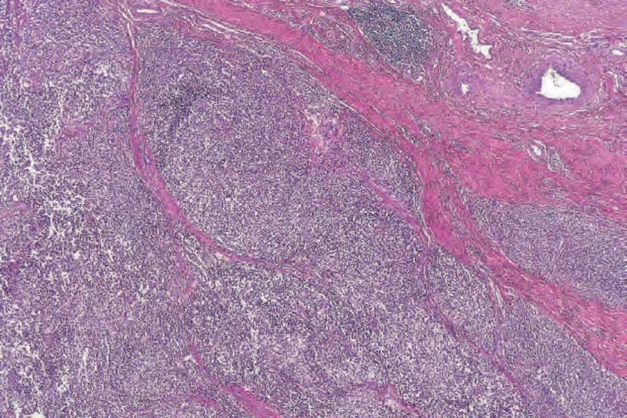
Fig. 35.405 Clear cell sarcoma: the tumor consists of nests of clear cells separated by fibrous septa.
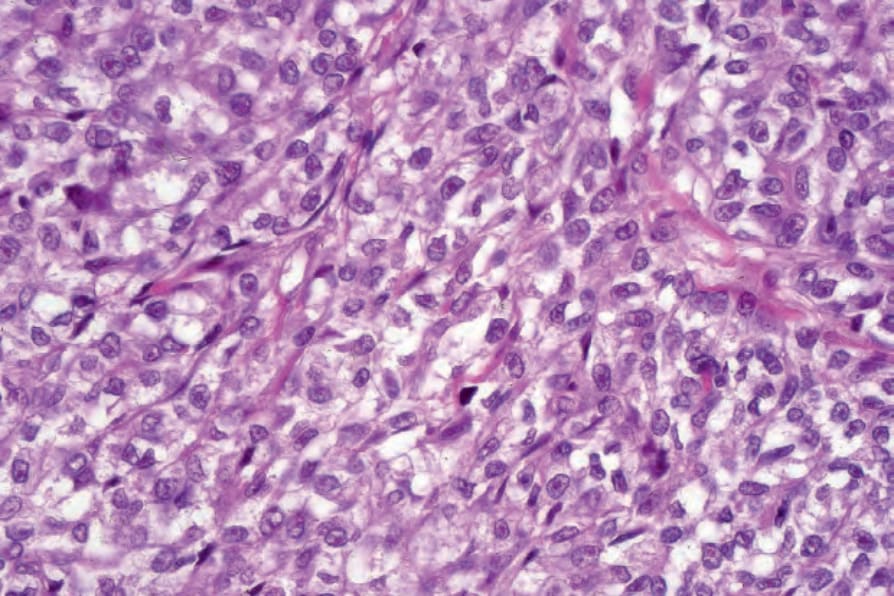
Fig. 35.406 Clear cell sarcoma: the tumor cells have clear cytoplasm and round vesicular nuclei with prominent eosinophilic nucleoli.
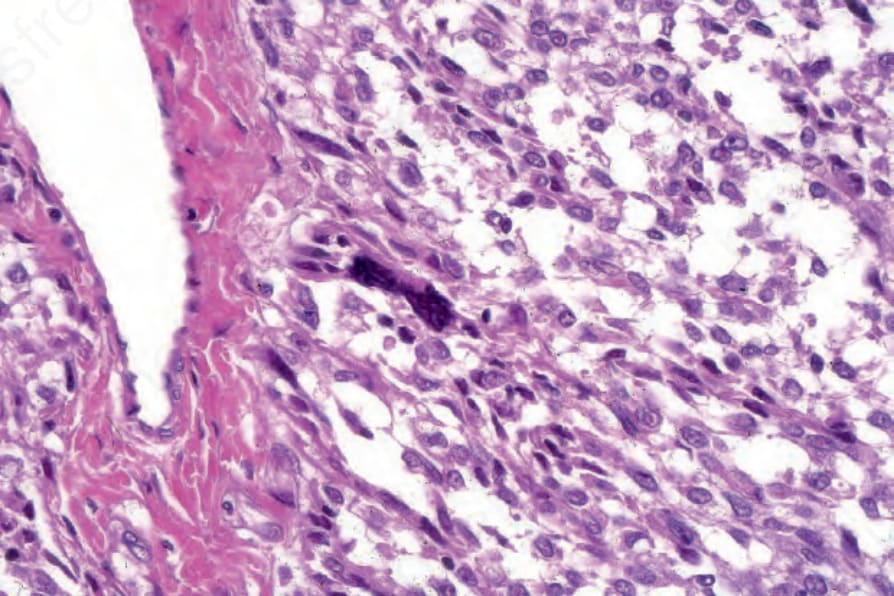
Fig. 35.407 Clear cell sarcoma: paraseptal irregular multinucleate cells are a common feature.
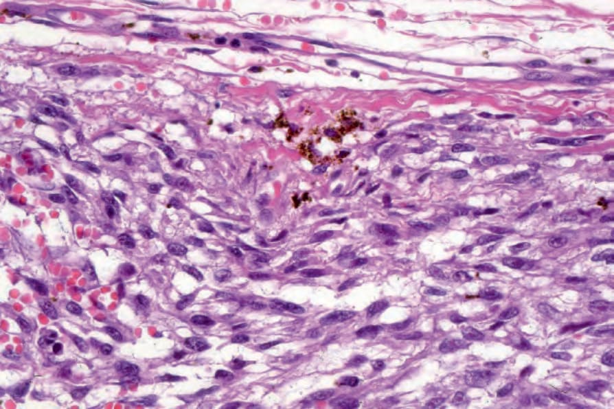
Fig. 35.408 Clear cell sarcoma: this section shows focal melanin pigmentation.