Glial heterotopias
Glial heterotopias
Clinical features Glial heterotopias are rare developmental congenital anomalies that are typically detected in infancy although very occasional cases are first noticed in adult life.1–5 In the latter setting, a case was detected because of visual loss.6 The majority are subcutaneous and present as a firm mass adjacent to the bridge of the nose, often with associated hypertelorism (Fig. 35.387). Up to one-third of cases are solely intranasal and a small proportion show both components. As most cases occur in or around the nose they are known as nasal gliomas. Lesions present as a nodule or polyp, and intranasal lesions often are accompanied by obstruction.7 However, rare examples can arise on the lip, pharynx, oral cavity (including the tongue), scalp and even the midline of the back and the sphenoid sinus.8–11 Multiple lesions are rare and a heterotopia and an encephalocele may occur simultaneously.12 A case associated with agenesis of the corpus callosum has been documented.13 A further case associated with PHACE syndrome (posterior fossa anomalies, hemangiomas of the face and scalp, arterial abnormalities, cardiac defects and eye anomalies) has also been reported.14
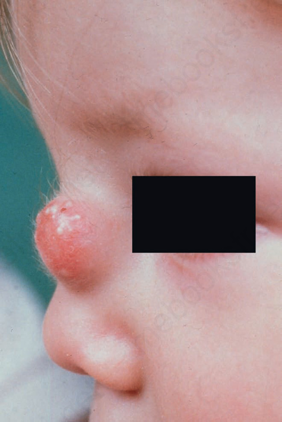
Fig. 35.387 Nasal glioma: the nose is the most commonly affected site. By courtesy of the Institute of Dermatology, London, UK.