Lipomatosis
Lipomatosis
Clinical features Lipomatosis is extremely rare and may present in several forms. Idiopathic forms include symmetric, diffuse and pelvic lipomatosis; the latter does not affect the superficial subcutaneous tissue.1–17
Two variants of lipomatosis affect the superficial subcutaneous tissue:
• Multiple symmetric lipomatosis (Launois-Bensaude) represents the commonest form and can be diffuse or localized.1–3,5 In the diffuse variant, there is usually symmetrical involvement of the trunk or proximal limbs, head and neck, pelvis, and occasionally the tongue, abdominal cavity and the intestinal tract;4,18 it is seen most often in children (particularly males), although adults may be affected. Single reports of associations with familial hyperlipidemia and tuberous sclerosis have been documented.16,17 Some cases show an autosomal dominant inheritance, others are associated with myoclonic epilepsy with ragged red fibers (MERRF), and a number may be associated with diabetes mellitus.6–9 The localized variant (multiple symmetric lipomatosis) is characteristically seen in the cervical region, back, shoulders and upper trunk, usually presents in middle-aged Mediterranean males with high alcohol intake, and is known as Madelung disease. Involvement of the mediastinum can be seen. Associated inspiratory dyspnea or obstructive sleep apnea may exceptionally occur.10,11 The axillae and groins can also be affected. There is strong association with alcohol abuse and liver disease.12 A variant of localized symmetrical lipomatosis restricted to the hands or the feet has been reported.13,14
• Asymmetric lipomatosis can present at any site and usually has no association with other diseases.15 Single reports of associations with familial hyperlipidemia and tuberous sclerosis have been documented.16,17
A localized form of lipomatosis of the scalp has been reported as encephalocraniocutaneous lipomatosis (Haberland disease) and is associated with alopecia, aplasia cutis, nevus psiloliparus, skin tags and ocular and cranial abnormalities.19–21 It is not associated with Proteus syndrome. A relationship with oculoectodermal syndrome has been proposed.21 Congenital facial infiltrating lipomatosis refers to a disorder associated with hypertrophy of bones and soft tissues, macrodontia and premature dental eruption.22,23
In addition, cases associated with exogenous or endogenous production of steroids (steroid lipomatosis) with predilection for the face, central chest, upper mid-back and spinal epidural, retro-orbital and mediastinal tissue may occur.24,25 An association with antiretroviral therapy (HIV lipodystrophy) has also been documented.26,27
In both idiopathic forms of lipomatosis, only radical surgery can prevent local recurrence. The disadvantages of recommending such treatment must be weighed against the possible functional impairment that the condition may induce.
Histologic features Mitochondrial DNA damage has been suggested as a possible etiology of symmetric lipomatosis and in lipodystrophy induced by HIV therapy.27,28 The localization of the lesions in multiple symmetric lipomatosis suggests an origin from brown fat.29 The demonstration that cells cultured from lesions synthetize mitochondrial inner membrane protein, a marker of brown fat, gives further support to this theory.29 Histologically, all forms are characterized by unencapsulated overgrowth of mature adipose tissue.
fibers and reduced numbers of epidermal appendages may also be a feature. Entrapped hair follicles may appear cystically dilated.
A case with focal pagetoid spread of adipocytes and dystrophic calcification has been described.18
As mentioned previously, it has been argued that the solitary form represents a pedunculated, fat-containing skin tag or a lipofibroma. Although the distribution of fat in the superficial dermis makes this suggestion unlikely, the argument is semantic and of no practical value. Nevus lipomatosus superficialis is also histologically indistinguishable from the cutaneous nodules of focal dermal hypoplasia.
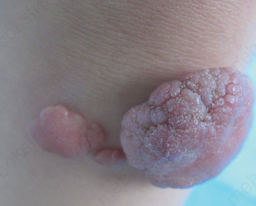
Fig. 35.12 Nevus lipomatosus superficialis: in this patient there are multiple papules and nodules. Courtesy of Dr Yi-Guo Feng, Xi’an Jiatong University, second affiliated hospital, China.
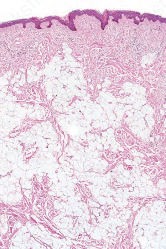
Fig. 35.13 Nevus lipomatosus superficialis: this specimen came from the lower back of a teenage male. There is widespread infiltration of the dermis by mature adipocytes.