Nevus lipomatosus superficialis (Hoffman and Zurhelle)
Nevus lipomatosus superficialis (Hoffman and Zurhelle)
Clinical features Nevus lipomatosus superficialis is an uncommon form of connective tissue nevus, manifest principally by the deposition of fatty tissue in the dermis.1–4 In its classical form, it is characterized by multiple papular, polypoid or plaque-like lesions, up to 2 cm in diameter, which almost always arise unilaterally on the posterior surfaces of the buttocks, upper thighs or lower back. More extensive and diffuse involvement may occur and patients present with prominent folds in what has been described as the Michelin tire appearance.5–7 Typically, the lesions present in early childhood or adolescence. Unusual associations include co-occurrence with lipedematous scalp, folliculosebaceous cystic hamartoma, dermoid cysts, angiokeratoma of Fordyce, perifollicular fibromas, and trichofolliculoma.8–13 A case associated with intramuscular lipomatosis has been reported.14 A solitary form, usually
seen in adults, shows a predilection for the same sites or occurs elsewhere and is more likely to represent a variant of fibroepithelial polyp or skin tag (Figs 35.11 and 35.12). Such lesions have been described as pedunculated lipofibroma.15 In all types of nevus lipomatosus the sex incidence is equal. The papules or plaques, varying from skin-colored to yellow, are characteristically broad based and may show superficial comedone formation.
Pathogenesis and histologic features The pathogenesis is unknown. In a single case, a 2p24 deletion has been described.16
Although alterations are seen in all the connective tissue elements in the dermis, the predominant feature is deposition of lobules of mature fat in variable quantities in the superficial dermis (Fig. 35.13).3,17 These fatty lobules are located particularly around small blood vessels, the numbers of which are also increased. Areas of loose fibrous tissue, diminished elastic
1703 Benign adipocytic tumors and tumorlike lesions
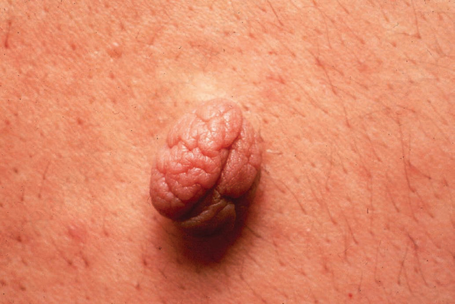
Fig. 35.11 Nevus lipomatosus superficialis: solitary lesions are often polypoid and have a soft consistency. By courtesy of the Institute of Dermatology, London, UK.
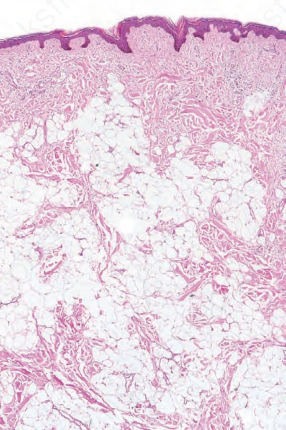
Fig. 35.13 Nevus lipomatosus superficialis: this specimen came from the lower back of a teenage male. There is widespread infiltration of the dermis by mature adipocytes.