Cutaneous metaplastic synovial cyst
Cutaneous metaplastic synovial cyst
Clinical features First described by González and coworkers in 1987, cutaneous metaplastic synovial cyst is an uncommonly reported lesion which usually follows surgical or other trauma.1–12 Patients present with an often tender dermal nodule adjacent to a scar and clinically diagnosed as a suture granuloma.1 Lesions draining serosanguinous fluid sometimes communicate with the surface epithelium. Exceptionally, multiple lesions may be encountered.7 Occasional examples have arisen without a history of trauma, most often in patients with severe rheumatoid arthritis.7,9 Two cases have presented in patients with Ehlers-Danlos syndrome.6,13 Lesions can occur at any site, and there is no sex or age predilection.7 Local recurrence is very rare.14
(Fig. 34.38).1,4 The core of the second type is composed of admixed spindled and epithelioid cells and a mixed inflammatory cell infiltrate in which multinucleate giant cells are sometimes conspicuous. The lining cells are devoid of atypia, but multiple (normal) mitoses are sometimes present. The cyst is surrounded by chronically inflamed granulation and scar tissue.3 Synovial metaplasia-like changes with papillary projections have rarely been described in association with oral mucoceles.21
The epithelioid cells regularly express vimentin and occasionally CD68, lysozyme, and α1-antichymotrypsin.1,2,9 They are consistently negative for keratins, CEA, EMA, S100 protein, SMA, and desmin.2,4,6,9
Pathogenesis and histologic features Seyle originally showed that a synovium-like membrane could develop in the connective tissue of rats following its disruption by the subcutaneous injection of air.15 The experiment was later repeated, and similar observations have been reported following implantation of various prosthetic devices.16–20
Cutaneous metaplastic synovial cyst is not a true cyst since it lacks an epithelial lining. It is located in the dermis underneath a sometimes thickened epidermis with which it occasionally communicates through a fistulous tract (Fig. 34.37).1 It contains multiple villous processes of two types: some are composed of hyalinized connective tissue covered by fibrin, whereas others are highly cellular and are lined by multilayered epithelioid cells
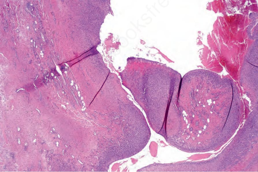
Fig. 34.37 Metaplastic synovial cyst: this example presented as a fistulous tract following abdominal surgery. Villous processes are evident.
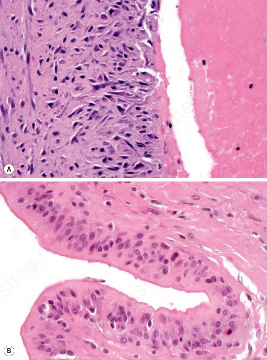
Fig. 34.38 (A, B) Metaplastic synovial cyst: the villi are covered by a layer of fibrin and vertically orientated spindle cells.