Branchial cyst
Branchial cyst
Cysts are unilocular or multilocular and contain clear, brown, red, or gelatinous material.4,5,9 The lining epithelium is variably stratified squamous, cuboidal, columnar, pseudostratified, or ciliated.1,3 Thymic remnants including Hassall corpuscles and cholesterol granulomata are also present.1,3,10 The adjacent fibrous capsule often contains lymphocytic aggregates.2 A lesion containing parathyroid tissue has been described.11
Clinical features The branchial (lymphoepithelial) cyst presents as a swelling near the angle of the jaw anterior to the sternomastoid muscle, most often at the junction of its upper one-third and lower two-thirds.1–4 Lesions also present in deeper tissues. The cyst is asymptomatic and does not move on swallowing.3 Patients are most commonly in their second or third decade. The sexes are affected equally.3 Rare lesions are associated with a fistule. A single case was associated with a duplicated facial nerve.5 Occasionally, bilateral cysts are present and then a familial tendency is sometimes in play.6
Pathogenesis and histologic features The origin of the cyst is uncertain although it is generally considered to represent a developmental anomaly of the branchial arches. Possibilities include incomplete obliteration of branchial mucosa, remnants of the precervical sinus, or an origin from the thymopharyngeal duct.3,7 Cystic degeneration of cervical lymph nodes has also been suggested.7
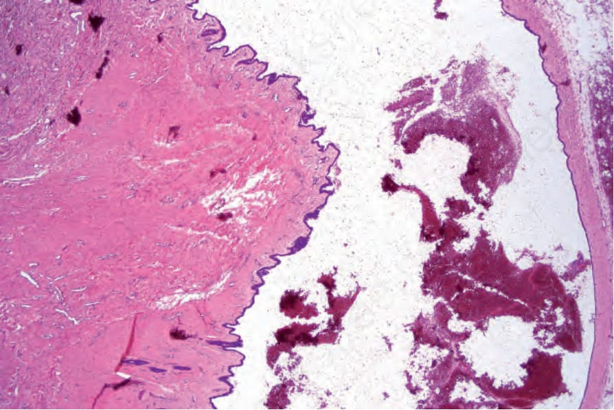
Fig. 34.33 Cutaneous ciliated cyst: note the papillary projections in the lower left of the field.