Trichofolliculoma
Trichofolliculoma
Clinical features Trichofolliculoma is a not uncommon hamartoma that usually arises on the face and presents as a single, dome-shaped, 0.5- to 1.0-cm-diameter papule with a central pore.1–7 It is occasionally found on the scalp or neck and has been rarely described on the vulva.7–9 A wide age range is affected, although lesions are very rare in children or infants.8,10,11 Characteristic, although not diagnostic, is the presence of one or more silky, white, thread-like hairs (trichoids) growing out of the central opening (Fig. 31.73).8 Rarely, trichofolliculoma may coexist with a basal cell carcinoma.12
Pathogenesis and histologic features It consists of a cystic cavity (dilated hair follicle) lined by stratified squamous epithelium (including a granular cell layer), which can usually be shown to arise from the surface epithelium (Fig. 31.74).1,2 The cavity contains keratinous debris and hair shaft fragments.8 Arising from its wall are numerous hair follicles, each surrounded by a clearly defined perifollicular sheath (Figs 31.75 and 31.76). Secondary budding with further abortive
1567 Folliculosebaceous cystic hamartoma
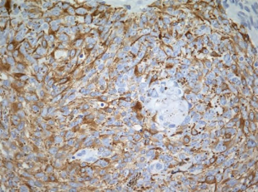
Fig. 31.72 Melanocytic matricoma: the dendritic melanocytes are highlighted with HMB-45.
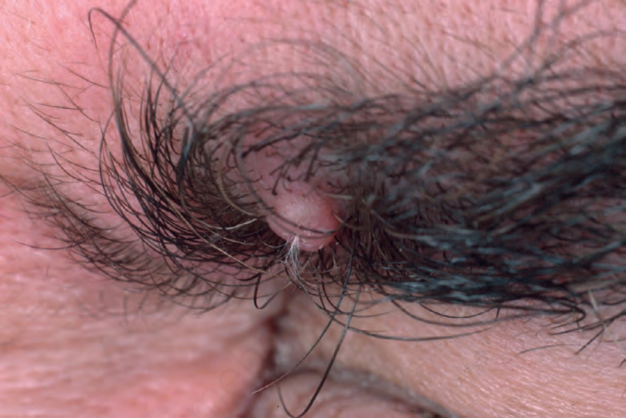
Fig. 31.73 Trichofolliculoma: characteristic dome-shaped nodule with protruding trichoids. From the collection of the late N.P. Smith, MD, the Institute of Dermatology, London, UK.

Fig. 31.74 Trichofolliculoma: scanning view showing a cystically dilated follicle communicating with the epidermis. Note the secondary follicles arising from the lateral wall.
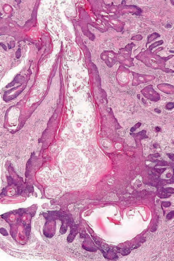
Fig. 31.75 Trichofolliculoma: this example emphasizes the numerous secondary follicles.
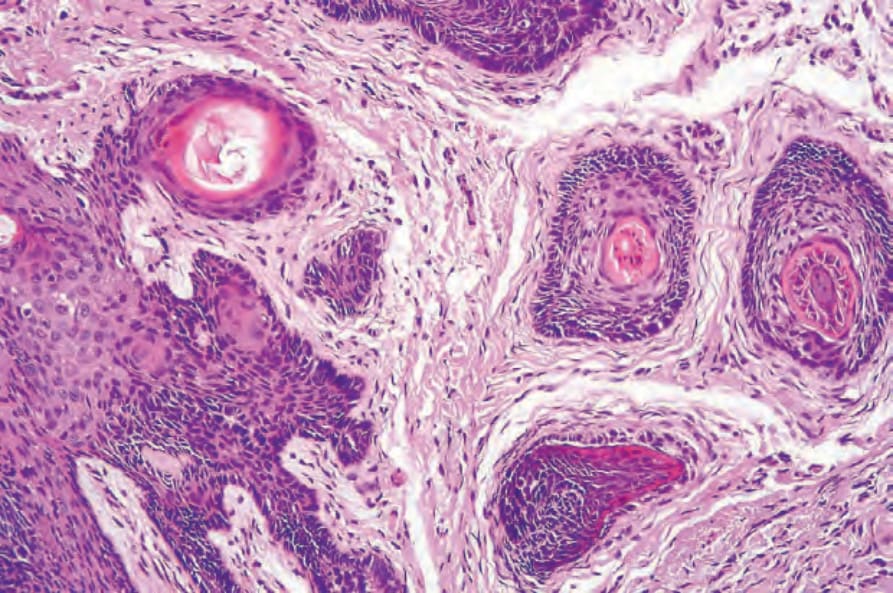
Fig. 31.76 Trichofolliculoma: high-power view of secondary follicles.