Trichoadenoma
Trichoadenoma
Clinical features Trichoadenoma is a rare tumor that most commonly presents on the face and to a lesser extent the buttocks of adults, occurring equally in men and in women.1–7 Very occasionally, the neck, upper arm, and thigh may be affected. Congenital and childhood presentation is unusual.4,8,9 The solitary asymptomatic lesions are soft or firm nodules 3–50 mm in diameter and variably yellowish or erythematous in color. They rarely present as linear and verrucous plaques.3,9 Development of a trichoadenoma within a dermal nevus is likely to be coincidental.10 Trichoadenoma is completely benign.
Histologic features Trichoadenoma is believed to be midway between trichoepithelioma and trichofolliculoma in terms of morphological differentiation and probably reflects differentiation toward the infundibular portion of the pilosebaceous canal.1,5,11
The epidermis is normal. Within the dermis is a well-defined fibroepithelial tumor composed of keratinous cysts and a conspicuous fibrovascular stroma (Fig. 31.17). The cyst wall is composed of squamous epithelium, which manifests epidermoid keratinization (with a granular cell layer) (Fig. 31.18). Solid epithelial islands may also sometimes be evident. There is generally no evidence of hair follicle formation, although in the verrucous variant (verrucous trichoadenoma), the cysts regularly contain vellus hairs.3,8
1551 Trichilemmoma and Cowden disease
Immunohistochemistry reveals retained CK20-positive intratumoral Merkel cells. Tumor cells are negative for Ber-EP4 and androgen receptor.12
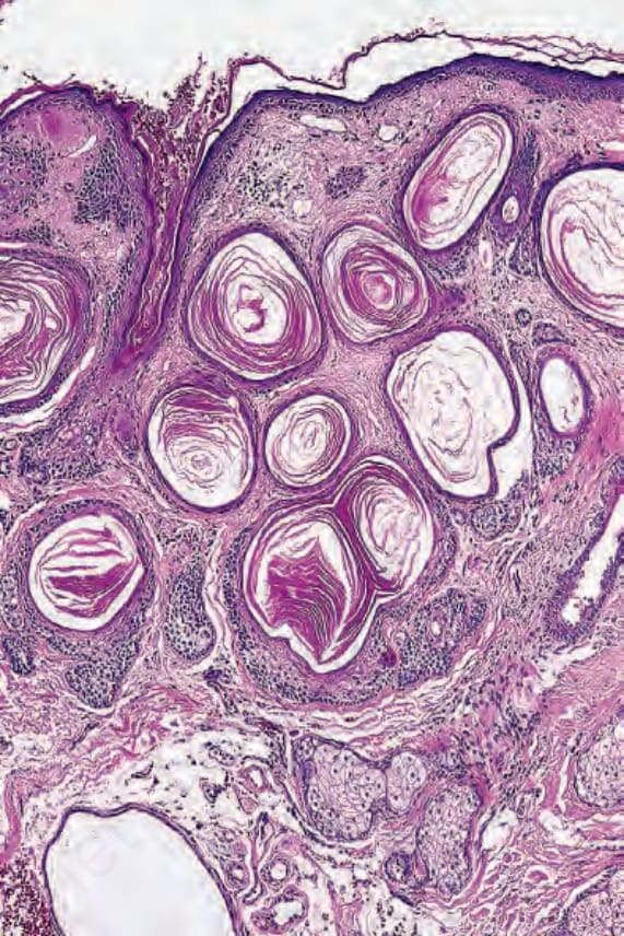
Fig. 31.17 Trichoadenoma: note the numerous keratocysts, many with a thick epithelial lining.
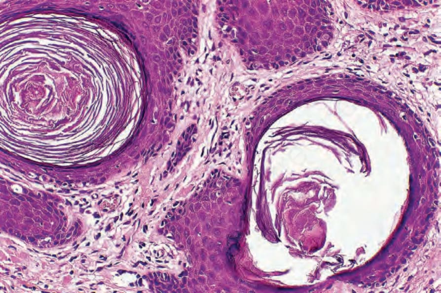
Fig. 31.18 Trichoadenoma: the cysts are lined by stratified squamous epithelium. Keratinization is epidermoid in type.