Maculopapular cutaneous mastocytosis/urticaria pigmentosa
Maculopapular cutaneous mastocytosis/urticaria pigmentosa
Maculopapular cutaneous mastocytosis is the commonest manifestation of mastocytosis, accounting for 80% of cutaneous cases, and with an approximate incidence of 1 : 1000–1 : 8000 live births.4,39,40 It presents most frequently in children, with no sex predilection.35 Lesions may present at birth or during the first year of life. They are pruritic, erythematous, or red-brown, round to oval macules, papules, and plaques, often measuring up to 2–3 cm in diameter (Figs 29.348 and 29.349).4,13 They occur predominantly on the trunk, although any region (including the mucous membranes) may be affected.24,40 Darier sign is characteristically positive and patients may show
1517 Mastocytosis
generalized dermographism. Blisters develop occasionally; rarely, these may be generalized and mimic a primary or acquired bullous dermatosis.42 In adults, lesions are more often disseminated, more heavily pigmented, and tend to be macular (Fig. 29.350).11 Telangiectasia may be present, hence the name, telangiectasia macularis eruptiva perstans.13,38,43,44
Most childhood cases resolve spontaneously before or during puberty. Adult cases tend to persist, and systemic mastocytosis should always be ruled out.1,11,12,38,45 Even when systemic mastocytosis is present, the course tends to be indolent and has no effect in survival.12
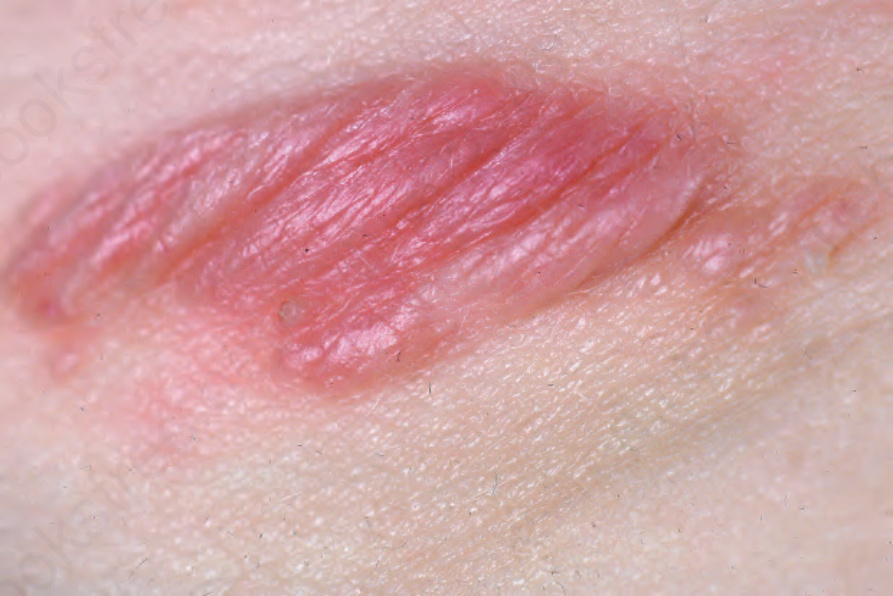
Fig. 29.347 Mastocytoma: edematous, erythematous plaque in a child. From the collection of the late N.P. Smith, MD, the Institute of Dermatology, London, UK.
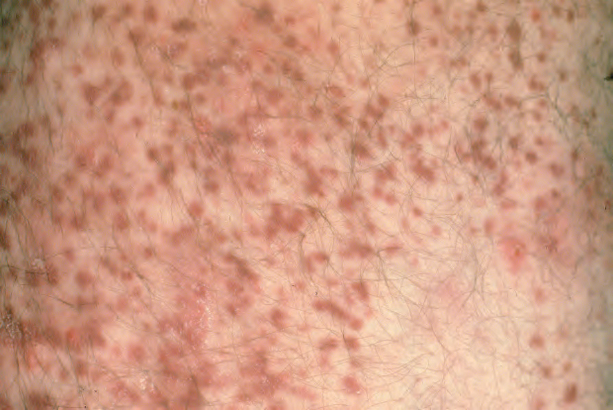
Fig. 29.348 Urticaria pigmentosa: numerous brown macules and papules are present. From the collection of the late N.P. Smith, MD, the Institute of Dermatology, London, UK.
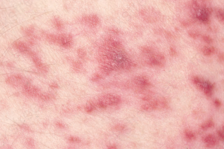
Fig. 29.349 Urticaria pigmentosa: in this example, the macules and papules are erythematous. From the collection of the late N.P. Smith, MD, the Institute of Dermatology, London, UK.
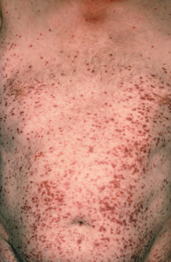
Fig. 29.350 Urticaria pigmentosa: note the generalized distribution of the lesions. From the collection of the late N.P. Smith, MD, the Institute of Dermatology, London, UK.