Mastocytosis
Mastocytosis
Mastocytosis was regarded as a myeloproliferative disorder in the 2008 WHO Classification of myeloid malignancies.1 However, major advances in the understanding of the disease has led to it now being categorized as a separate disease entity in the 2016 update of the WHO classification.2 It is a heterogeneous disorder, characterized by clonal proliferation and accumulation of mast cells in one or more organ systems. The clinical course ranges from asymptomatic, sometimes spontaneously regressing disease with a normal life expectancy, to highly aggressive forms associated with multiorgan failure and short survival.1,3–5 Mastocytosis is subdivided into different categories on the basis of extent and distribution of organ involvement, degree of impairment of organ function, and other clinical and laboratory findings.1–9 The two main subtypes are systemic mastocytosis (20% of cases), and a more common, skin-limited form of the disease referred to as cutaneous mastocytosis (80% of cases). Systemic mastocytosis is characterized by involvement of bone marrow and/or other extracutaneous organs, although skin may also be involved in up to 50% of cases. Diagnosis of systemic mastocytosis requires fulfillment of at least one major and one minor, or three minor criteria (Table 29.6).
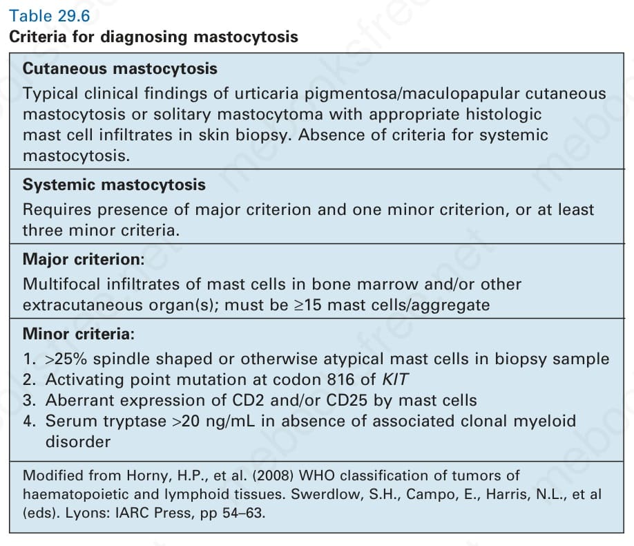
Table 29.6 Criteria for diagnosing mastocytosis