Benign cephalic histiocytosis
Benign cephalic histiocytosis
Benign cephalic histiocytosis is exceptional and presents in early childhood.33–41 The age at onset ranges from 3 to 34 months (mean, 13.5 months).34 The sexes are equally affected. Early lesions are erythematous, round or oval maculopapules that enlarge to form 2–8 mm diameter, brownish-yellow papules distributed most often on the face, particularly the cheeks, eyebrows, and forehead (Fig. 29.297). Not infrequently, they later spread to affect the shoulders, proximal limbs, trunk, and pubic area.34,35
Numbers of lesions are variable, ranging from solitary to hundreds of papules.34 The mucous membrane, palms, and soles are unaffected and there is generally no evidence of systemic involvement. An exceptional case with diabetes insipidus and a further case with insulin-dependent diabetes mellitus have been reported.40,42 Spontaneous regression always occurs, leaving transitory hyperpigmented lesions, which subsequently disappear completely.
1499 Xanthogranuloma family
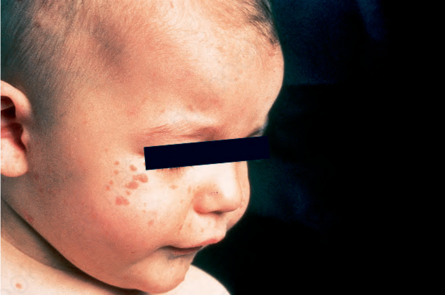
Fig. 29.297 Benign cephalic histiocytosis: numerous erythematous macules and papules are present on this infant’s forehead and cheek. By courtesy of R. Gianotti, MD, Universitá di Milano, Milan, Italy.
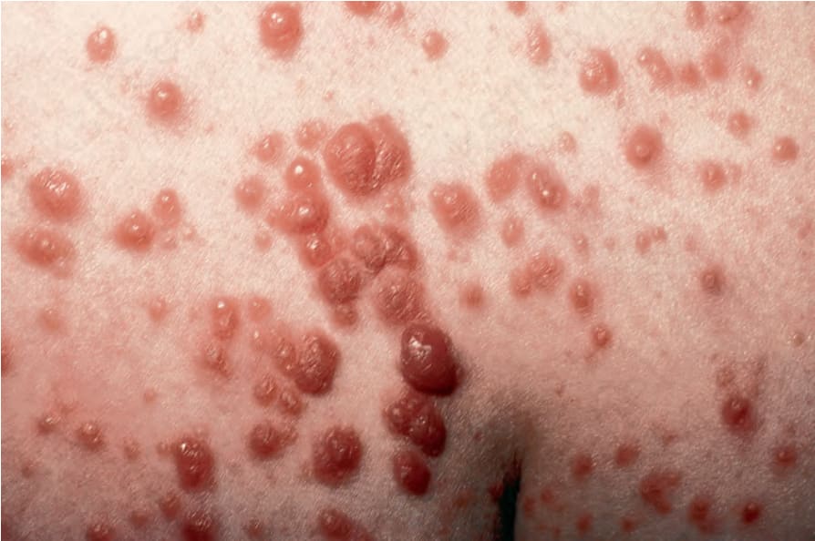
Fig. 29.298 Generalized eruptive histiocytoma: this middle-aged patient developed hundreds of asymptomatic papules on the trunk and limbs. By courtesy of R.M. Mackie, MD, Western Infirmary, Glasgow, UK.
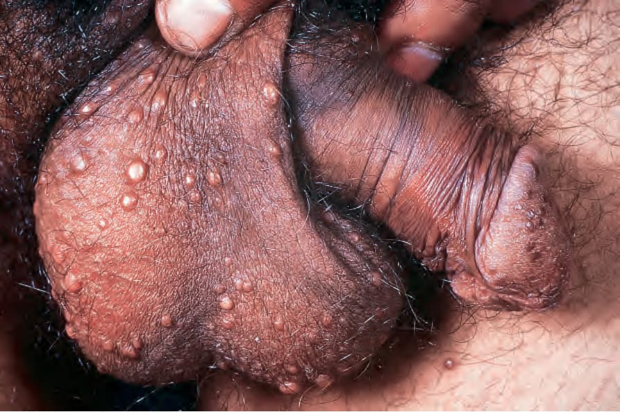
Fig. 29.299 Xanthoma disseminatum: typical papules on the scrotum. By courtesy of the Institute of Dermatology, London, UK.
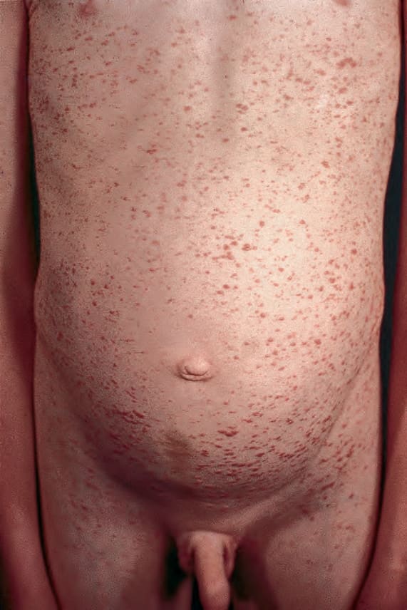
Fig. 29.300 Xanthoma disseminatum: in this patient, lesions are generalized. By courtesy of the Institute of Dermatology, London, UK.