Amyloidosis
Amyloidosis
Clinical features Conjunctival amyloidosis is an uncommon condition that is usually localized and rarely associated with systemic involvement. It is usually seen in middle-aged adults. Patients present with confluent fusiform lesions or polypoid papules with a waxy or yellow color. Some patients may present with subconjunctival hemorrhage (Fig. 27.57) or blepharoptosis. Conjunctival amyloidosis usually affects the palpebral conjunctiva, but any part of the conjunctiva may be affected. Systemic evaluation for primary systemic amyloidosis and lymphoma is warranted.2,3
Histologic features Conjunctival amyloidosis is characterized by homogeneous eosinophilic deposits in the conjunctival substantia propria that are Congo red and thioflavin T positive (Fig. 27.58A and B). With Congo red and polarized light, the deposits show apple-green birefringence and dichroism (Fig. 27.58C).
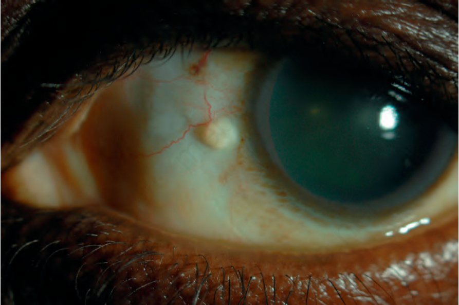
Fig. 27.56 Pinguecula: a yellow-white mass is seen on the nasal bulbar conjunctiva.
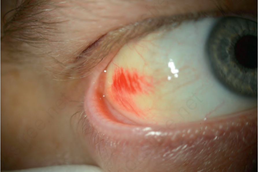
Fig. 27.57 Conjunctival amyloidosis: a yellow, waxy lesion with overlying subconjunctival hemorrhage is seen on the temporal bulbar conjunctiva.