Neurofibroma
Neurofibroma
Clinical features Neurofibroma is a peripheral nerve tumor that occurs rarely in the conjunctiva. It may be solitary, plexiform, or diffuse. Plexiform neurofibroma is associated with neurofibromatosis type 1. Solitary neurofibroma is usually not associated with systemic disease. It presents as a slow-growing, nodular, amelanotic epibulbar mass in adults.1–3 Plexiform neurofibroma is diffuse.
Histologic features Neurofibroma is composed of Schwann cells and fibroblasts in a fibromyxoid matrix. Solitary neurofibromas may be circumscribed, but not encapsulated. It consists of spindle cells with elongated, slender, sometimes wavy nuclei (Fig. 27.52). Scattered mast cells and nerve axons are present. By immunohistochemistry, S100 protein is generally positive throughout most of the tumor. CD34 highlights perineural fibroblasts, and neurofilament highlights axons within the lesion.
Clinical features Granular cell tumor is rare in the conjunctiva and caruncle.1 It appears as a pink to red stromal mass that may be clinically indistinguishable from other well-circumscribed tumors.2
Histologic features Granular cell tumor is composed of sheets of large polygonal cells with small, bland-appearing nuclei and abundant granular eosinophilic cytoplasm. The granular cytoplasm is PAS positive and diastase resistant. There may be overlying pseudoepitheliomatous hyperplasia. By immunohistochemistry, tumor cells are positive for S100 protein and neuron-specific enolase.
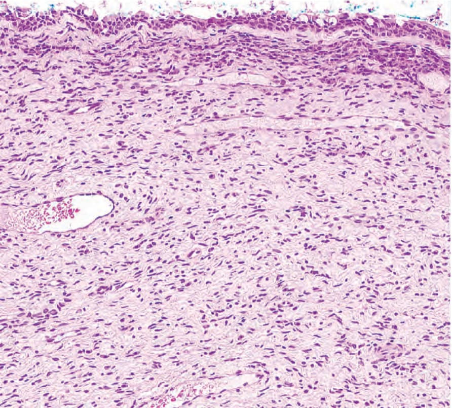
Fig. 27.52 Neurofibroma: the tumor is composed of cells with eosinophilic cytoplasm and wavy nuclei.