Primary dermal melanoma
Primary dermal melanoma
Clinical features The described cases of this apparently rare presentation of melanoma show a dermal nodule lacking an identifiable in situ component or association with a pre-existing nevus.1–4 Published series indicate that this presentation of melanoma represents less than 1% of all melanomas.5–7 A wide anatomic distribution is reported, but the head and neck region and extremities are the most common sites. In one study, in a cohort of 13 patients, the average Breslow depth was 9.6 mm with 92% survival at a mean of about 4 years.8,9 Subsequent studies have documented a somewhat less impressive but still relatively favorable 88% 5-year survival in a cohort of nine cases with a median Breslow thickness of 3.4 mm.6 A series of 49 cases with mean Breslow depth of 3.0 mm and mean mitotic rate of 4.3 per mm2 is also reported.7 A caveat of this latter series is that 14 cases showed association with a pre-existing intradermal nevus and average follow-up was only 26 months. Nineteen percent of patients developed locoregional recurrence and 6% showed distant metastases; only one patient died of disease. Traditional AJCC parameters did not correlate with outcome. These studies suggest that this tumor behaves much less aggressively than comparable traditional nodular melanoma. Others examining a larger series of 101 cases defined as a solitary dermal melanoma (as distinct from melanoma of unknown primary which usually presents as an isolated lymph node metastasis) within larger well-characterized cohort of 12,817 patients found no difference in outcome compared to stage-matched traditional melanomas.5 Other studies show various results, but most include melanoma of unknown primary which could skew the results.10–14 Indeed, in all of these papers, it is unclear whether the authors are describing the same tumor. Much more study is needed to refine our definitions and understanding of this possible entity and delineate its natural history. At the present time, this presentation is probably best assessed and reported as a standard melanoma with risk proportional to the assessed Breslow depth, though a note suggesting that such cases have been described to have more indolent behavior would certainly be appropriate.
Histologic features The lesion is characterized by a nodular deposit of melanoma composed of epithelioid to spindled cells in the dermis and/or subcutis. Spitzoid and blue nevus-like features have been noted in some cases, but many have the appearance of traditional melanoma.7 Some cases have conspicuous mitoses, but overall a Ki-67 index may be lower than that seen in comparable cases of nodular melanoma or metastatic melanoma.8 It would be extremely important to distinguish these cases from metastatic melanoma or melanoma of unknown primary, both of which have vastly different staging implications.15 This can be challenging and indeed such cases have now been described to have BRAF and NF1 mutations as well as P16/ CDKN2A loss and other copy number aberrations similar to traditional cutaneous melanoma.7,16 Explanations for this entity include an origin in association with follicular melanocytes and complete regression of a prior intraepidermal component. More recently, de la Fouchadiere and colleagues have described five cases of non-pigmented dermal melanoma each with a CRTC1-TRIM11 fusion that are dermal based and nodular in outline.17 Cytology ranges from epithelioid to spindled sometimes with scattered multinucleate cells. In some ways the appearance is similar to that of clear cell sarcoma which can also sometimes be confined to the dermis.18 Behavior is low grade. This lesion would appear to fall within the spectrum of primary
dermal melanoma though where it falls in the end in the spectrum between melanoma and dermal clear cell sarcoma remains to be seen.
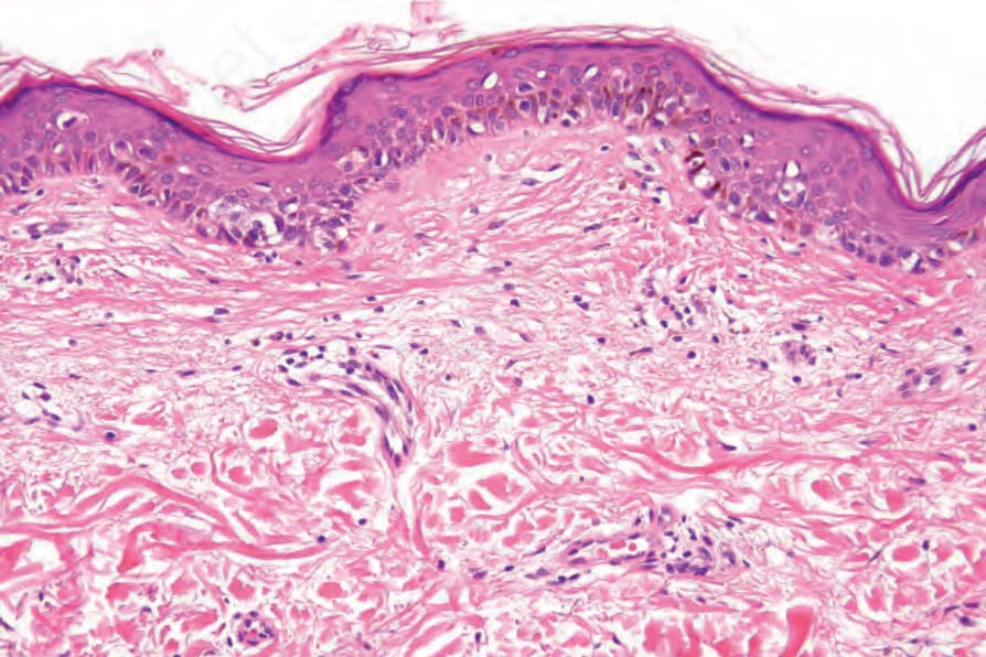
Fig. 26.132 Lentiginous melanoma: the tumor cells are predominantly basally located. There is no evidence of solar elastosis.
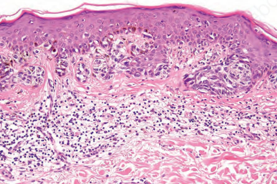
Fig. 26.133 Lentiginous melanoma: junctional nests showing moderate to severe cytological atypia are evident.