Lentiginous melanoma
Lentiginous melanoma
Clinical features This type of melanoma should be distinguished from acral lentiginous melanoma. These tumors have a propensity for the shoulders and upper back in elderly patients while the lower legs, particularly in younger female patients, may also be affected.1–6 Similar to lentigo maligna, this form of melanoma may be associated with an extended in situ phase of many years, and in an important number of cases there is a precursor lesion. The latter have been described as atypical lentiginous melanocytic nevus and dysplastic atypical nevus of the elderly with some questioning whether these lesions are truly malignant.7–9 Additional study is required to further define and characterize this seemingly distinct lesion.
Histologic features Lentiginous melanoma, as the name implies, consists of a primarily single cell array of melanocytes achieving confluence at least focally with limited associated junctional nests (Figs 26.131–26.133). Pagetoid intraepidermal ascent can be noted but it tends to be very focal when present. Epidermal atrophy is not present and there is preservation of normal rete architecture
1354 Melanoma
at the dermal–epidermal junction. Cytological atypia varies and may not be prominent, and solar elastosis is not a feature. These features help separate this entity from other forms of lentiginous melanoma. Melanocytic stains may be helpful to better visualize the increased density of atypical melanocytes. These lesions may be more readily recognized in larger excision specimens as the features may be difficult to discern in smaller biopsies.2 As stated before, in many lesions there is evidence of a lentiginous nevus with variable atypia, suggesting a precursor lesion. This is the reason the diagnosis can be missed in small biopsies.

Fig. 26.128 Basomelanocytic tumor: surrounding undifferentiated tumor cells is a striking palisade of basophilic cells typical of basal cell carcinoma. By courtesy of L. Erickson, MD, Mayo Clinic, Rochester, Minnesota.
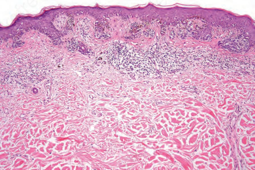
Fig. 26.131 Lentiginous melanoma: in this field, there is a lentiginous and focally nested atypical melanocytic proliferation. Note the upper dermal lymphocytic infiltrate.