Blue nevus-like melanoma (malignant blue nevus)
Blue nevus-like melanoma (malignant blue nevus)
This term covers a variety of lesions including melanoma arising in a background of cellular blue nevus and to a lesser extent common blue nevus or showing morphological overlap with blue nevus.1–5 Variants have also been documented complicating nevi of Ito and Ota and pilar neurocristic hamartoma.6–8 It also includes cases of melanoma that show histologic overlap with cellular blue nevi but which are devoid of a precursor lesion (de novo variants).9–13 Pigmented epithelioid melanocytoma (previously termed animal-type or pigment-synthesizing melanoma; see below) can have a highly similar morphological spectrum but are distinct from blue nevus-like melanoma at the genomic level.14
Clinical features Malignant blue nevi are often very slowly growing lesions that commonly present as a consequence of a sudden onset of growth and show a predilection for the scalp (Fig. 26.80). Males are affected more often than females. No age group is immune and exceptionally children are affected. This is a high-grade lesion with outcome similar to stage-matched cases of traditional melanoma cases.15,16 The lung, liver, and lymph nodes are most commonly affected by metastatic disease.
Histologic features Tumors that arise in a background of pre-existent blue nevus typically show an abrupt transition from a benign precursor lesion to obvious melanoma (Figs 26.81 and 26.82). The latter presents as one or more nodules of
epithelioid or spindled melanocytes showing a diffuse growth pattern and obvious cytological features of malignancy (Figs 26.83 and 26.84).
Tumors that mimic cellular blue nevus, but in which no precursor lesion can be identified, commonly show an expansile growth pattern with usually pushing borders extending into the subcutaneous fat (Fig. 26.85). Occasionally a dumbbell scanning morphology may be evident and rarely an alveolar growth pattern is seen.11 Typically, at low-power magnification, a diagnosis of benign cellular nevus is anticipated. The tumor cells most often have spindled cell morphology but epithelioid and mixed variants are sometimes encountered. High-power detail, however, shows an increased nuclear-to-cytoplasmic ratio, mild to moderate nuclear pleomorphism, hyperchromatism, nucleolar prominence, and increased mitotic activity (Figs 26.86 and 26.87). Necrosis, sometimes exhibiting a geographic pattern, may also be present and perineural infiltration is an occasional feature.
1340 Melanoma
A
B
Scattered dendritic cells are often present in this second variant. Whether this represents a residual benign precursor lesion or indicates malignant dendritic cells is problematical. In favor of the latter possibility is the presence of dendritic cells in metastases (Fig. 26.88).
A subset of melanocytic proliferations is difficult to classify definitively as benign or malignant. These have been referred to as atypical cellular blue nevi or cellular blue melanocytic proliferation of uncertain malignant potential.1,17 There appears to be a lack of consensus regarding diagnostic criteria for malignancy across the blue nevus spectrum.18
Both blue nevus and blue nevus-like melanoma are associated with mutation in GNAQ or less commonly GNA11, encoding the alpha subunit of a heterotrimeric G-protein.14,19 Similar to ocular melanoma, mutations in BAP1, SF3B1, and EIF1AX are commonly encountered in blue nevus-like melanoma and their presence may help to establish malignancy.14,20 The standard multi-probe FISH assay can be helpful in distinguishing blue
1341 Histologic variants of melanoma
A
B
nevus-like melanoma from atypical forms of cellular blue nevus, but limited data are available.21,22
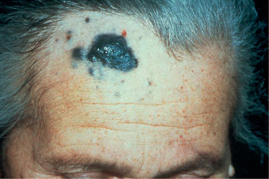
Fig. 26.80 Malignant blue nevus: note the heavily pigmented primary tumor associated with multiple satellite lesions on this elderly patient’s forehead. By courtesy of the Institute of Dermatology, London, UK.
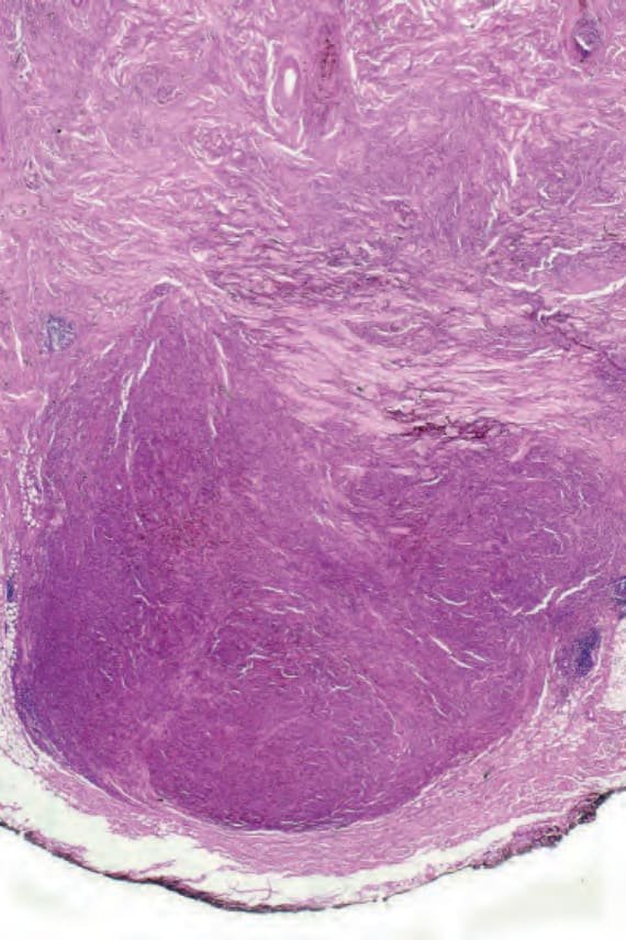
Fig. 26.81 Malignant blue nevus: this tumor arose on the scalp. There is a dense expansile tumor nodule which has extended into the subcutaneous fat.
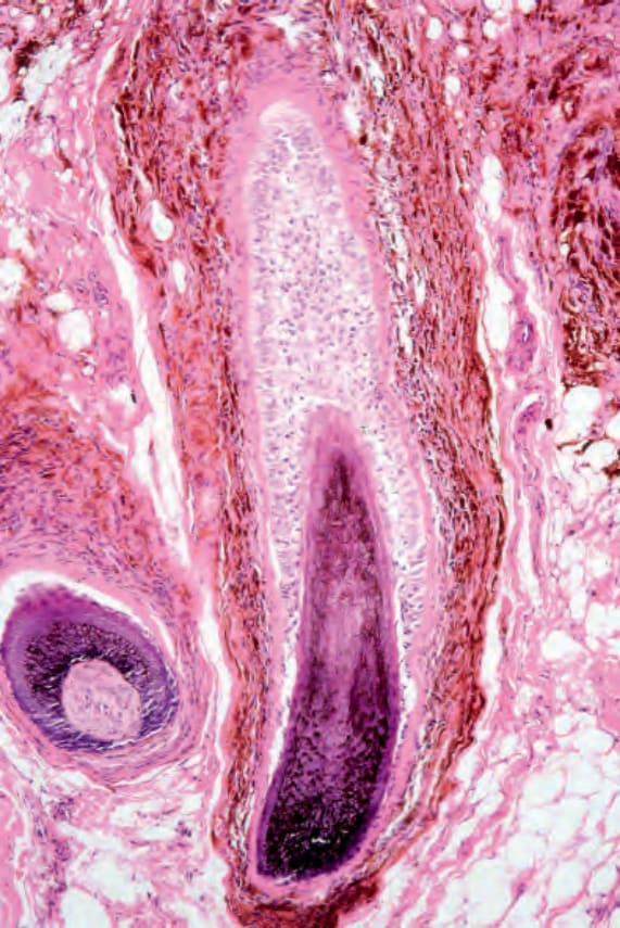
Fig. 26.82 Malignant blue nevus: a pilar blue nevus was evident in the adjacent skin.
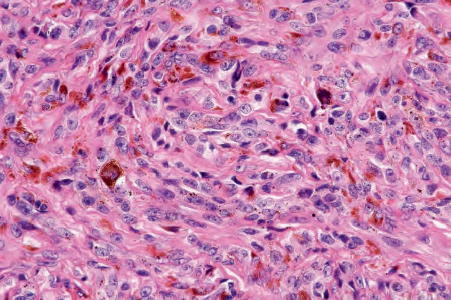
Fig. 26.83 Malignant blue nevus: the tumor cells have vesicular nuclei with prominent nucleoli.
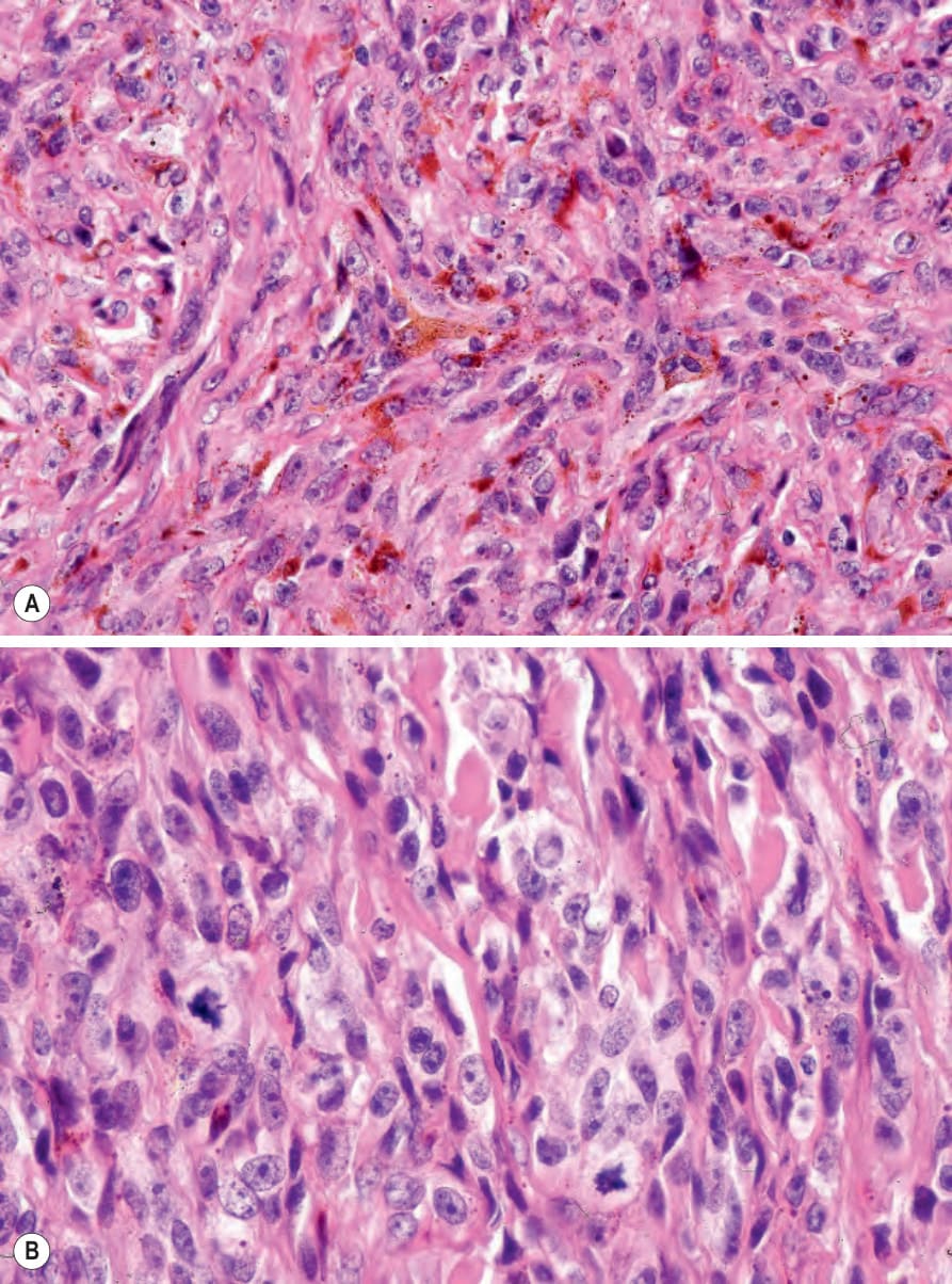
Fig. 26.84 Malignant blue nevus: (A) there are multiple dendritic cells; (B) note the two mitoses.
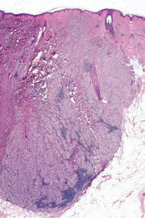
Fig. 26.85 Malignant blue nevus: a precursor lesion was not identified in this case.
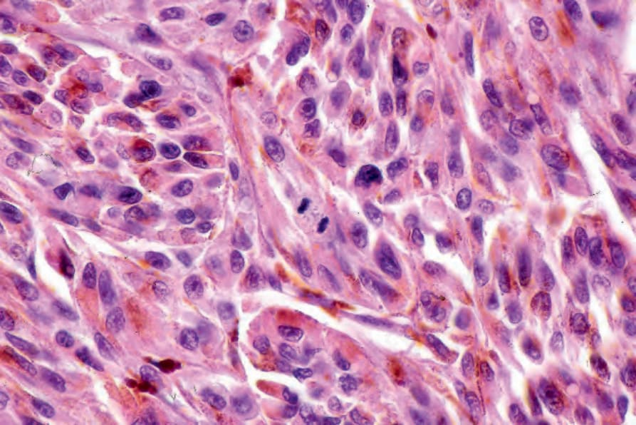
Fig. 26.86 Malignant blue nevus: note the central mitotic figure.
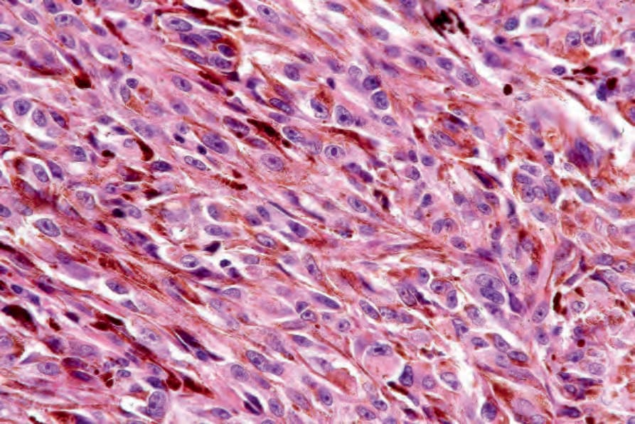
Fig. 26.87 Malignant blue nevus: dendritic cells are conspicuous in this field.
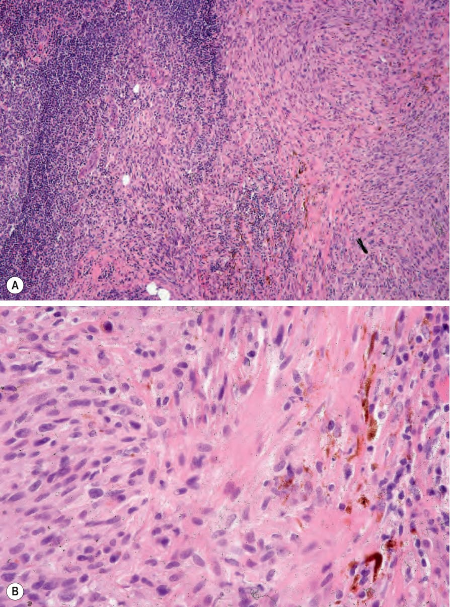
Fig. 26.88 (A, B) Malignant blue nevus: the sentinel node contained metastatic melanoma. A small number of dendritic cells are present. This is the same case as illustrated in Figures 26.81–26.84.