Melanoma with neuroendocrine differentiation
Melanoma with neuroendocrine differentiation
Neuroendocrine differentiation has been rarely described in melanoma cases and sometimes in non-cutaneous cases.1–4 Mixed melanocytic and neuroendocrine tumors have also been described in the lung, thymus, and thyroid and challenge current classification schemes.5 Immunohistochemical expression of chromogranin and synaptophysin, along with neurofilament protein, has been noted as has expression of CD56.3 Less specific markers of neuroendocrine differentiation such as neuron-specific enolase and perhaps CD56 expressed in isolation should be interpreted with caution when seen in melanoma as they may not imply such differentiation. Characteristic melanocytic markers remain intact. While not clearly associated with any difference in natural history, neuroendocrine differentiation can be associated with diagnostic confusion, especially with presentation at sites where other neuroendocrine tumors can be seen such as in the sinonasal region.6 Further complicating the distinction is that melanoma exhibiting
1339 Histologic variants of melanoma
neuroendocrine differentiation appears to show small cell morphology. Interestingly, carcinoid-like trabecular and pseudorosette patterns have been noted in melanoma as well, but these cases lacked neuroendocrine markers.7,8
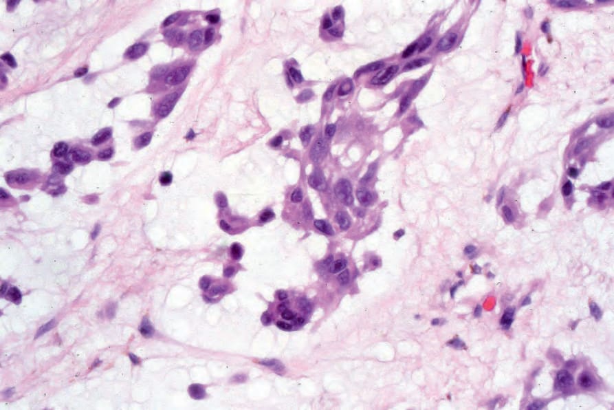
Fig. 26.79 Adenoid (pseudoglandular) melanoma: high-power view showing undifferentiated tumor cells dispersed within abundant mucin. By courtesy of R. Margolis, MD, St Elizabeth’s Medical Center, Boston, USA.
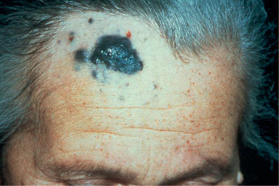
Fig. 26.80 Malignant blue nevus: note the heavily pigmented primary tumor associated with multiple satellite lesions on this elderly patient’s forehead. By courtesy of the Institute of Dermatology, London, UK.