Myxoid melanoma
Myxoid melanoma
Myxoid stromal change in melanoma is very rare and may be seen in primary, recurrent, and metastatic disease.1–11 It is of no prognostic significance.4,8
Myxoid change is a feature of vertical growth phase melanoma. It may be evident as a minor component of the tumor or be present throughout. Tumors showing the latter can be a source of considerable diagnostic difficulty with epithelioid cases suggesting carcinoma and spindle cell cases mimicking sarcoma. It has been described as a reactive change after phototherapy.12,13 Typically, the tumor cells are smaller than those present in nonmyxoid areas and may be epithelioid, spindled, or stellate in appearance (Fig. 26.77). Melanin pigmentation is variable. The mucin, which presents as basophilic ‘stringy’ material, is usually PAS negative, colloidal iron and Alcian blue positive, and sensitive to hyaluronidase (consistent with stromal mucin).8 Often the melanoma cells are present as discohesive clumps, cords, and strands such that a pseudoglandular or ‘acantholytic’ appearance may sometimes result (Figs 26.78 and 26.79). Occasionally, pseudoacinar differentiation and intercellular molding reminiscent of adenocarcinoma are features.8
Immunohistochemistry is typical for melanoma, the tumor cells being invariably positive for S100 protein and usually positive for HMB-45.8 HMB-45 negativity has, however, been documented.5
Myxoid change may sometimes be seen in the stroma of desmoplastic melanoma, but other areas more typical of conventional desmoplastic melanoma are usually present.
Myxoid melanoma may be confused with mucus-secreting adenocarcinoma and myxoid malignant peripheral nerve sheath tumor (MPNST) or other sarcomas. The former may be excluded on the basis of negative keratin immunocytochemistry and absence of PAS-positive mucin. Myxoid malignant peripheral nerve sheath tumor usually shows much less strong and only focal S100 protein expression, and HMB-45 is negative. In addition, MPNST limited to the dermis is exceedingly rare.
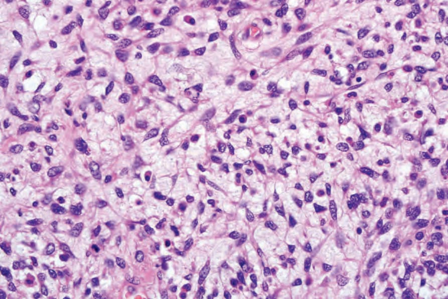
Fig. 26.77 Myxoid melanoma: this rare variant can be a source of considerable diagnostic difficulty. Note the ‘undifferentiated’ irregular cells associated with an abundant myxoid stroma.
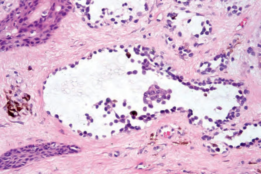
Fig. 26.78 Adenoid (pseudoglandular) melanoma: this variant also associated with excess mucin deposition may easily be misdiagnosed as an acantholytic squamous carcinoma or adenocarcinoma if a junctional component or pigment is absent. Diagnosis often depends upon immunohistochemistry. By courtesy of R. Margolis, MD, St Elizabeth’s Medical Center, Boston, USA.