Balloon and clear cell melanoma
Balloon and clear cell melanoma
Balloon cell melanoma is a very rare vertical growth phase variant with less than 20 cases well documented in the medical literature.1–5 There are no particular distinguishing clinical features except that a polypoid configuration
Balloon cells have abundant eosinophilic or clear cytoplasm showing fine granularity or vacuolation (Figs 26.74 and 26.75). The tumor cells are typically S100 protein, Mart-1, SOX10, and HMB-45 positive. They contain PAS-positive, diastase-resistant granules. These are also iron hematoxylin positive, hyaluronidase resistant. Sensitivity to ribonuclease indicates that the granules are composed of ribonucleoprotein.2 The cause of the vacuolation is uncertain but is believed to represent either abnormal melanosome metabolism or else a melanosome degenerative phenomenon.
Balloon cell melanoma can be distinguished from balloon cell nevus by the absence of nuclear pleomorphism and mitotic activity in the latter.7 Balloon cell melanoma may also be confused with sebaceous and xanthomatous tumors. Sebaceous lesions have a characteristic ‘bubbly’ cytoplasm and often the nucleus is crenated. In addition, sebaceous tumors express epithelial membrane antigen as well as adipophylin and are S100 protein and HMB-45 negative. Xanthomatous lesions contain lipid, express CD68, and are S100 protein negative.7–10
1338 Melanoma
Pure clear cell variants due to intracytoplasmic glycogen deposition are very rare and may be encountered in primary tumors or metastatic deposits (Fig. 26.76).5,11–16 They may be confused with clear cell carcinomas, including clear cell squamous carcinoma, clear cell hidradenocarcinoma, and clear cell metastases such as from clear cell carcinoma of the kidney. In cases where there is real diagnostic difficulty, the use of immunocytochemistry for melanocytic and keratin expression will resolve the issue.
Exceptionally, multiple small intracytoplasmic vacuoles can give rise to a pseudolipoblast appearance with conspicuous nuclear scalloping.
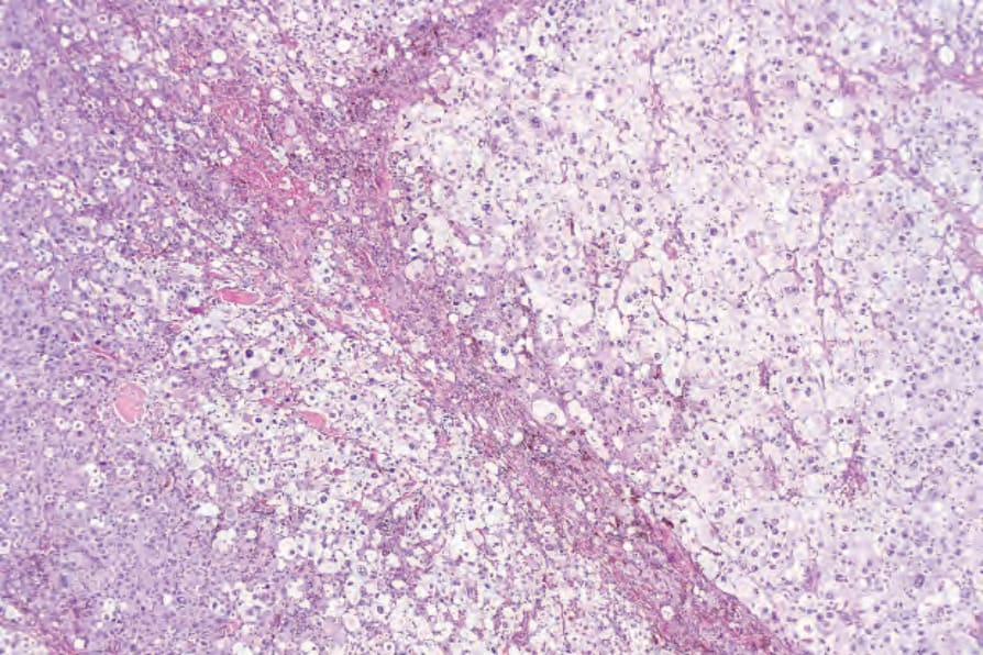
Fig. 26.74 Balloon cell melanoma: the tumor cells have clear or faintly granular cytoplasm. This example is amelanotic and could easily be mistaken for a xanthomatous lesion.
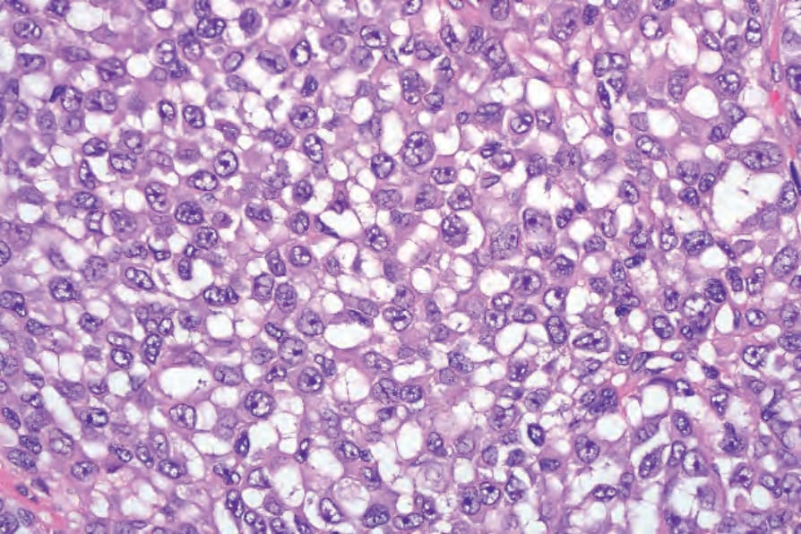
Fig. 26.76 Clear cell melanoma: this is an exceedingly rare variant of melanoma. In the absence of a known history of melanoma or visible melanin pigment, the diagnosis depends upon immunohistochemistry.
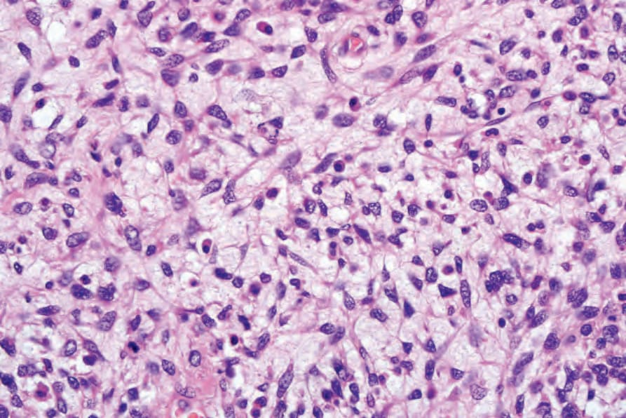
Fig. 26.77 Myxoid melanoma: this rare variant can be a source of considerable diagnostic difficulty. Note the ‘undifferentiated’ irregular cells associated with an abundant myxoid stroma.