Minimal deviation melanoma
Minimal deviation melanoma
The concept of minimal deviation melanoma was introduced to define a subset of invasive melanoma that showed minimal histologic deviation from banal nevi and therefore lacked the pleomorphism seen in classic melanoma. It was further postulated that such tumors appeared to have a better prognosis than classic melanoma.1–8 Over the last decade, use of this nomenclature has precipitously declined and it is now presented primarily for historical interest.
Immunohistochemistry for the BRAF V600E mutation using the VE1 clone is a highly sensitive and specific means of determining the mutational status of melanoma, usually applied in the metastatic setting.109–111 It does not reliably detect common alternative BRAF V600 variants such as V600K that constitute 15–20% of BRAF mutations in some series.112,113 This can be helpful in the rapid assessment for eligibility for BRAF inhibitor therapy, but DNA sequencing is still the gold standard for determining BRAF mutational status. A similar approach can detect a subset of the common NRAS mutant proteins Q61R and Q61L in melanoma with two separate antibody clones, but is not widely adopted.110,114–118
In the setting of immunotherapy, membranous expression of PD-L1 (programmed death-ligand 1) can influence the choice of immuno-oncological agents, but is not required to use such approaches.119–122 A number of antibodies are available against PD-L1 for use in routine clinical immunohistochemistry,
As defined, two subtypes were recognized6:
• borderline minimal deviation melanoma where the dermal component is confined to the papillary dermis (Clark level 3),
• minimal deviation melanoma where there is infiltration into the reticular dermis or beyond (Clark level 4 or 5). Minimal deviation melanoma is characterized by the presence of an expansile nodule showing only mild to moderate cytological atypia. It lacks the disorderly, more marked pleomorphism of conventional epithelioid or spindled cell melanoma. The spectrum includes epithelioid variants composed of cells resembling those of a compound melanocytic nevus, spitzoid forms, spindled cell variants, and halo nevus-like lesions.1
1331 Histologic variants of melanoma
Although widely publicized, the concept has not gained general acceptance.1 This relates particularly to a lack of precise definitions combined with the well-known unpredictable behavior of many melanomas. Patients with apparently high-grade and thick tumors sometimes survive for many years, or even decades, while the converse may also be true. The postulated good behavior of these lesions has been challenged on the basis that those that neither recurred nor metastasized may have been benign nevi and those that metastasized and/or proved fatal were obviously malignant at the outset.
In our view, although there are histologic variants of melanoma that show only limited morphological deviation from banal nevi, the evidence relating to biological behavior suggests that these tumors fare no better than classical melanoma and should be treated in exactly the same way. Subtypes that could be included within this category of ‘minimal deviation’ are nevoid melanoma, small cell melanoma, and spitzoid melanoma. We prefer these categories for histologic classification and these three are dealt with below.
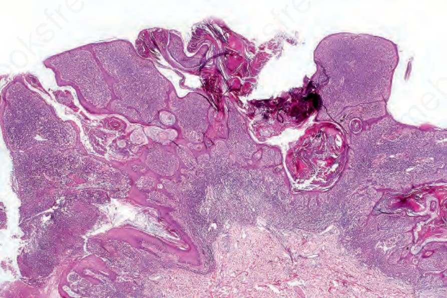
Fig. 26.57 Verrucous nevoid melanoma: verrucous nevoid lesions in the elderly should be viewed with suspicion.
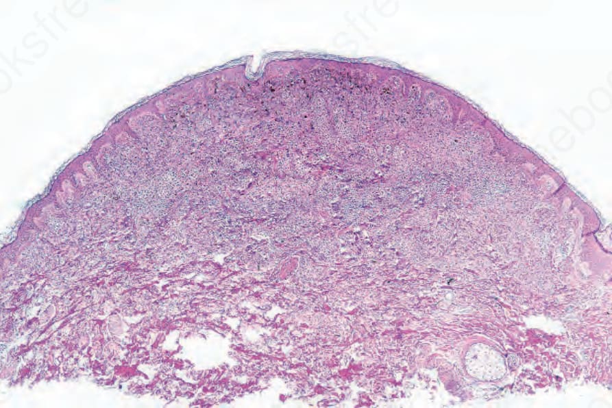
Fig. 26.58 Nodular nevoid melanoma: the diagnosis of nodular nevoid melanoma depends upon careful scrutiny of all ‘nevi.’ Inspection at scanning magnification will ensure misdiagnosis as a banal nevus!