Superficial spreading melanoma (pagetoid melanoma)
Superficial spreading melanoma (pagetoid melanoma)
The radial growth phase of superficial spreading melanoma is now encountered more commonly, largely as a consequence of public awareness of melanoma with the consequent removal of an increasing percentage of thin early lesions. It is typified by an asymmetrical proliferation of atypical nondendritic melanocytes scattered singly and in clusters throughout all levels of the epithelium (buckshot scatter), giving an appearance reminiscent of Paget disease (Figs 26.27 and 26.28). The melanoma cells involve all layers
of the epithelium while the cells of Paget disease tend to be superficial to the basal cell layer. The individual cells are epithelioid with abundant cytoplasm, often showing fine or dusty melanin pigmentation, and contain pleomorphic vesicular nuclei with prominent eosinophilic nucleoli; scattered mitotic figures including atypical forms may be evident. Characteristic of this variant is tumor growth in continuity from one rete ridge to another (a pattern not usually seen in acquired junctional or compound nevi). In contrast to lentigo maligna, there is often minimal visible evidence of actinic damage.159 The epidermis often shows acanthosis with partial or complete effacement of the ridge pattern. Invasive tumor is mostly of the epithelioid type (Figs 26.29–26.32). Desmoplasia and/or neurotropism are uncommon.
1318 Melanoma
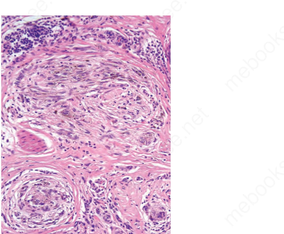
Fig. 26.23 Lentigo maligna melanoma: higher-power view.
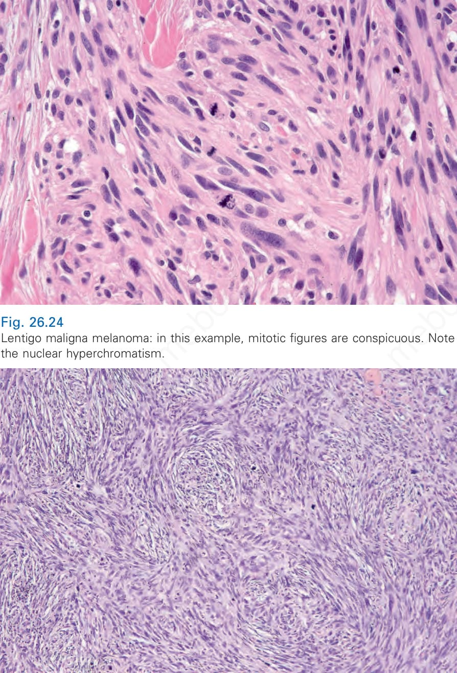
Fig. 26.24 Lentigo maligna melanoma: in this example, mitotic figures are conspicuous. Note the nuclear hyperchromatism.
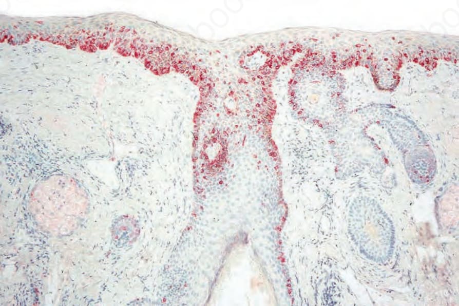
Fig. 26.25 Lentigo maligna melanoma: occasionally the tumor adopts a storiform pattern. When amelanotic, this may be confused with dermatofibrosarcoma protuberans or spindle cell squamous carcinoma.
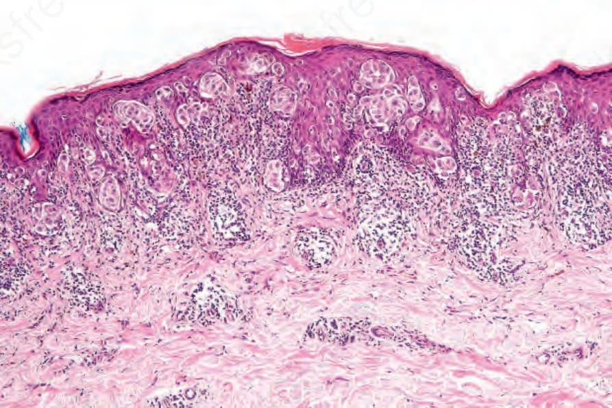
Fig. 26.27 Superficial spreading melanoma: there is prominent junctional activity and atypical melanocytes are widely scattered throughout the epidermis.
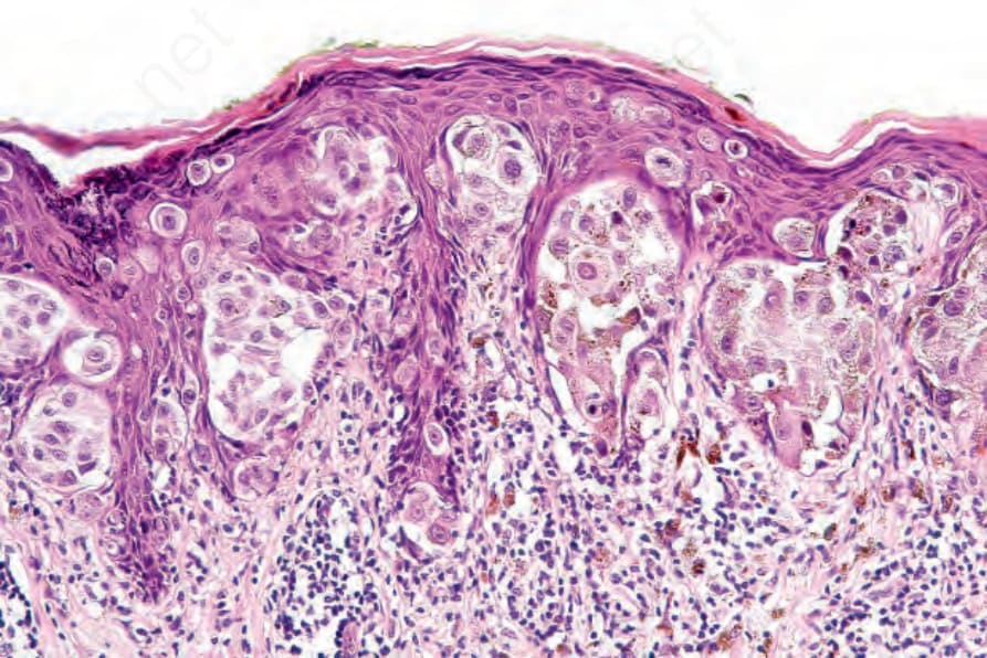
Fig. 26.28 Superficial spreading melanoma: the melanocytes have abundant eosinophilic cytoplasm and pleomorphic vesicular nuclei. Nucleoli are conspicuous.
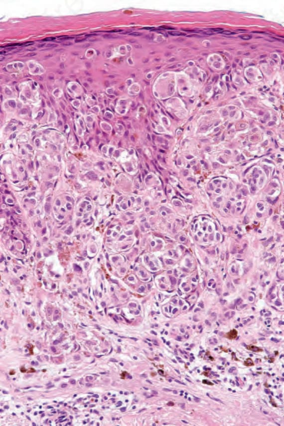
Fig. 26.29 Superficial spreading melanoma: invasive tumor is usually of the epithelioid type as shown in this field. Note the abundant cytoplasm, nuclear pleomorphism, and prominent nucleoli.