Acral lentiginous melanoma
Acral lentiginous melanoma
Acral lentiginous melanoma accounts for approximately 8–10% of all melanomas in Caucasians.123,124 It is, however, the predominant subtype affecting Afro-Caribbeans and Asians.125–127 The tumor is particularly found on the digits (especially beneath the nails) and on weight-bearing sites; plantar tumors are the most common, with the heel being the most frequently affected region (Figs 26.7 and 26.8).128,129 Subungual variants, which most commonly affect the great toe and the thumb, are rare tumors, accounting for only 2% of all cutaneous melanomas. They show a female preponderance and present most often in the elderly.123 In addition to acral lentiginous lesions, subungual melanoma can also present as superficial spreading and nodular histologic variants.130–132
Acral lentiginous tumors usually present as irregular, gradually enlarging, and variably pigmented macules. With progression to vertical growth phase,
1313 Histologic features
frequently ulcerated, blue or black nodular lesions are encountered. In Caucasians, acral lentiginous melanoma presents most often in the seventh decade, has an equal incidence in both sexes, and is generally associated with a poor prognosis since tumors are generally thick by the time of diagnosis. Mucosal melanomas are often classified within the acral lentiginous spectrum, given certain partial morphological overlap. These two types of melanoma are distinct clinically, although genomic characterization does indicate shared features.103,133,134
for a vascular tumor, such as lobular capillary hemangioma (pyogenic granuloma) (Figs 26.9 and 26.10).
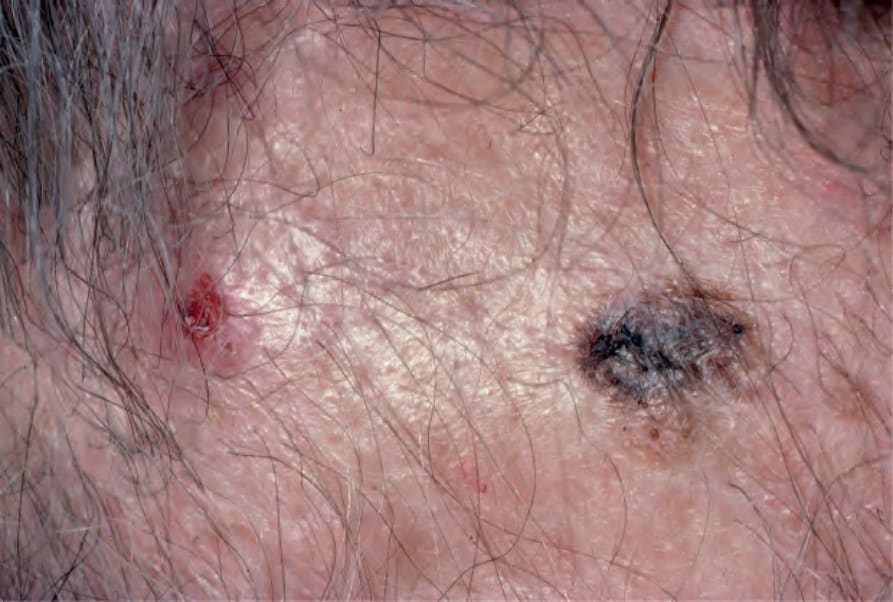
Fig. 26.5 Lentigo maligna: close-up view. From the collection of the late N.P. Smith MD, the Institute of Dermatology, London, UK.
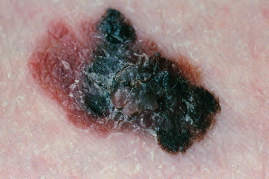
Fig. 26.6 Superficial spreading melanoma: there is a large nodule of invasive melanoma with a surrounding macular component. From the collection of the late N.P. Smith MD, the Institute of Dermatology, London, UK.
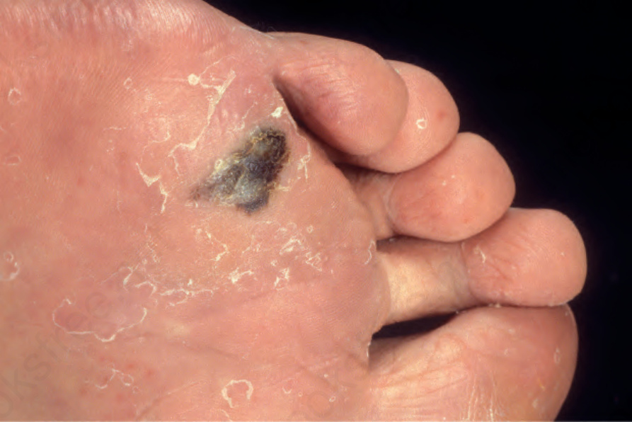
Fig. 26.7 Acral lentiginous melanoma: this variant arises on the non–sun-damaged skin of the palms, soles, and under the nails. From the collection of the late N.P. Smith MD, the Institute of Dermatology, London, UK.
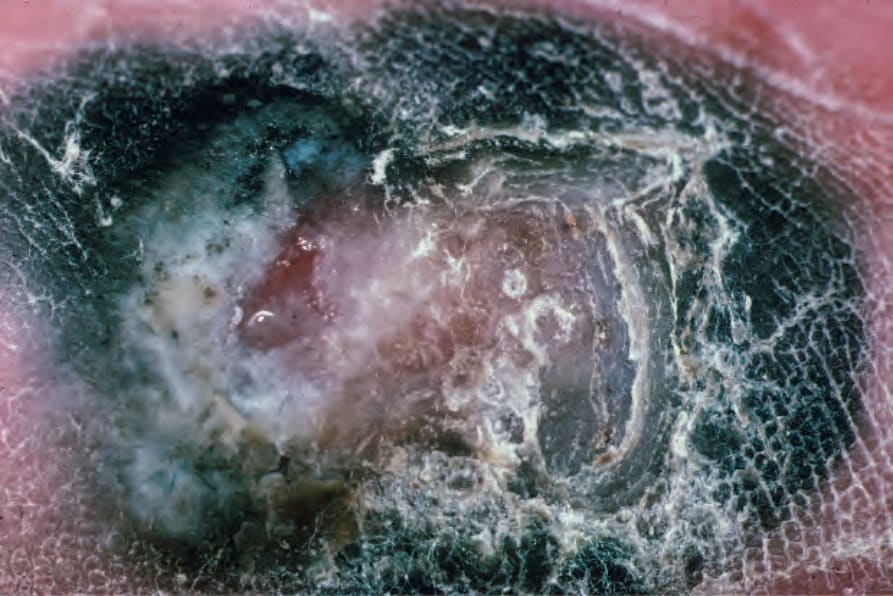
Fig. 26.8 Acral lentiginous melanoma: this ulcerated tumor had extended deeply into the underlying tissues. From the collection of the late N.P. Smith MD, the Institute of Dermatology, London, UK.
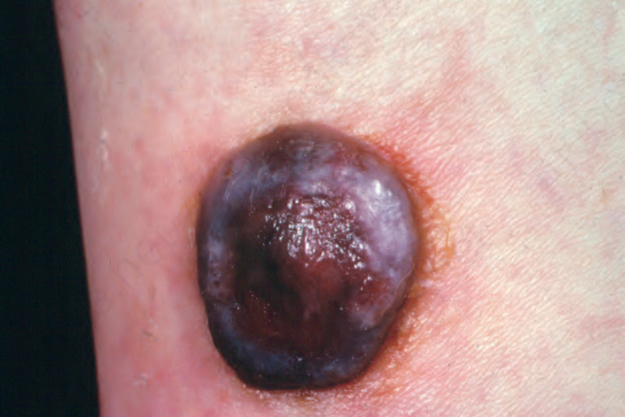
Fig. 26.9 Nodular melanoma: this heavily pigmented, dome-shaped nodule has no adjacent macular component. From the collection of the late N.P. Smith MD, the Institute of Dermatology, London, UK.