Lentigo maligna and lentigo maligna melanoma
Lentigo maligna and lentigo maligna melanoma
Lentigo maligna (Hutchinson melanotic freckle) is a relatively uncommon variant of melanoma and typically develops on chronic sun-damaged skin of the elderly.115–117 In the United States, it is estimated to account for 4% of all cases of cutaneous melanoma.118 It is characterized by a positive correlation
1312 Melanoma
with increasing age.119,120 Sites of predilection are the malar region, nose, temple, and forehead; much less frequently acral sites, such as the dorsum of the hands, may be involved (Figs 26.1–26.5). The tumor presents most often in the sixth and seventh decades as a variably pigmented, gradually enlarging, irregular, flat macule. It may be brown or black and usually shows areas of hypopigmentation representing areas of regression. The in situ lesion is often present for 10–15 years before invasive tumor develops. Lentigo maligna melanoma is a term used to denote to lentigo maligna that has progressed to dermal invasion.121
border of the lesion is characteristic, and hypopigmented areas of regression are also a frequent finding. Ulceration is an important diagnostic (and prognostic) clue.
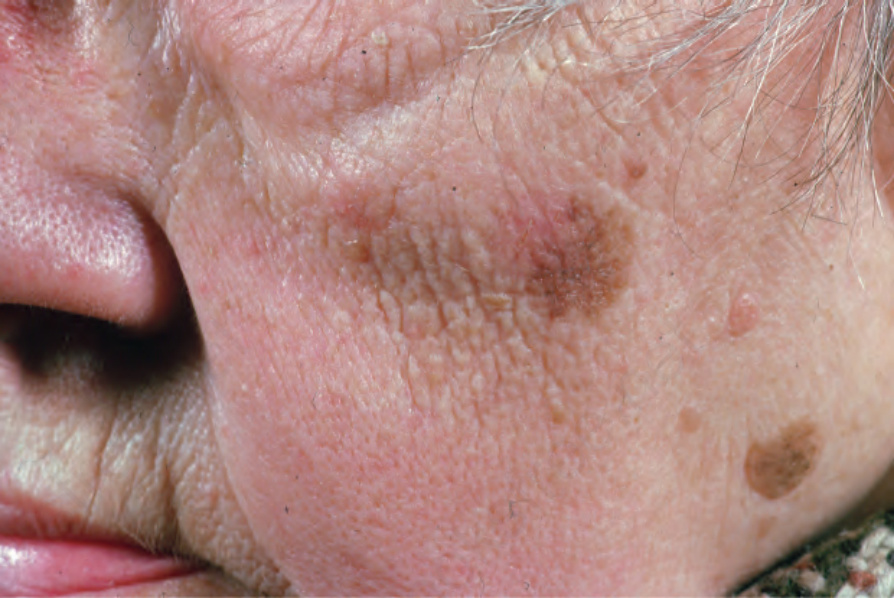
Fig. 26.1 Lentigo maligna: this variably pigmented lesion was present for many years. There is no invasive component. From the collection of the late N.P. Smith MD, the Institute of Dermatology, London, UK.
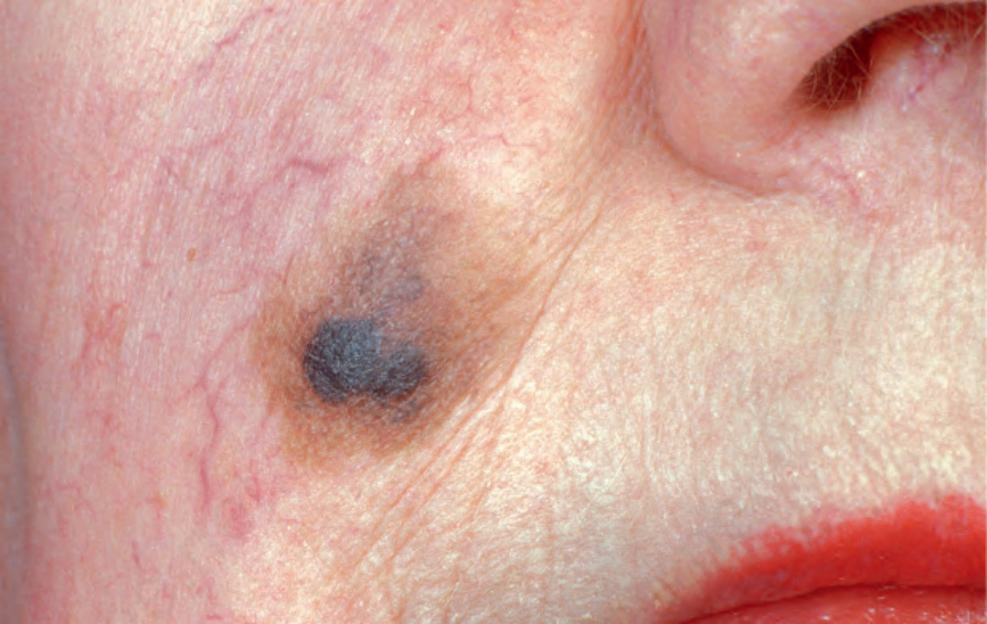
Fig. 26.2 Lentigo maligna: a dark black nodule of invasive tumor is surrounded by typical lentigo maligna. From the collection of the late N.P. Smith MD, the Institute of Dermatology, London, UK.
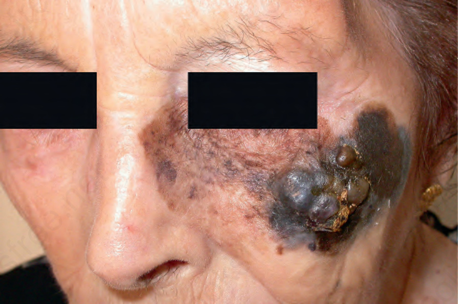
Fig. 26.3 Lentigo maligna melanoma: this is a very advanced lesion with a multinodular invasive component. By courtesy of J.C. Pascual, MD, Alicante, Spain.
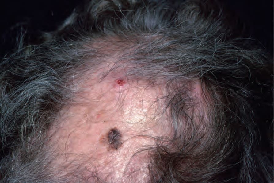
Fig. 26.4 Lentigo maligna: the sun-damaged skin of the bald scalp is at risk for developing this tumor. A concomitant ulcerated basal cell carcinoma is also evident above the melanoma. From the collection of the late N.P. Smith MD, the Institute of Dermatology, London, UK.